

294
JOHN RUNNSTRÖM
(Α)
(Β)
(Ο
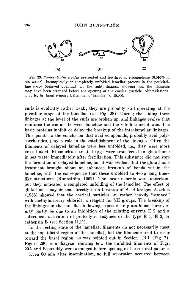
FIG. 29. Paracentrotus lividus pretreated and fertilized in ribonuclease (0.016% in
sea water). Incompletely or completely unfolded lamellae present in the perivitelline space (delayed opening). To the right, diagram showing how the filaments
may have been arranged before the opening of the cortical particle. Abbreviations:
c, curls; br, basal region; 1, filament of lamella. X 15,000.
curls is evidently rather weak; they are probably still operating at the
pivotlike stage of the lamellae (see Fig. 28). During the sliding these
linkages at the level of the curls are broken up, and linkages evolve that
reinforce the contact between lamellae and the vitelline membrane. The
basic proteins inhibit or delay the breakup of the intralamellar linkages.
This points to the conclusion that acid compounds, probably acid polysaccharides, play a role in the establishment of the linkages. Often the
filaments of delayed lamellae were less unfolded, i.e., they were more
cross-linked. Ribonuclease-treated eggs were transferred to glutathione
in sea water immediately after fertilization. This substance did not stop
the formation of delayed lamellae, but it was evident that the glutathione
treatment brought about an enhanced breakup of bonds within the
lamellae, with the consequence that these unfolded to 4-5 μ long fiberlike structures (Runnström, 1962). The measurements were uncertain,
but they indicated a completed unfolding of the lamellae. The effect of
glutathione may depend directly on a breakup of S—S bridges. Afzelius
(1956) showed that the cortical particles are rather heavily "stained"
with methylmercury chloride, a reagent for SH groups. The breakup of
the linkages in the lamellae following exposure to glutathione, however,
may partly be due to an inhibition of the gelating enzyme Ε 2 and a
subsequent activation of proteolytic enzymes of the type Ε 1, Ε 3, or
cathepsin Β (see Section II,D).
In the resting state of the lamellae, filaments do not necessarily meet
at the top (distal region of the lamella), but the filaments tend to recur
toward the basal region, as was pointed out in Section Ι,Β,Ι (Fig. 7).
Figure 29C is a diagram showing how the unfolded filaments of Figs.
29A and Β possibly were arranged before opening of the cortical particle.
Even 60 min after insemination, no full separation occurred between
JOHN RUNNSTRÖM
(Α)
(Β)
(Ο
FIG. 29. Paracentrotus lividus pretreated and fertilized in ribonuclease (0.016% in
sea water). Incompletely or completely unfolded lamellae present in the perivitelline space (delayed opening). To the right, diagram showing how the filaments
may have been arranged before the opening of the cortical particle. Abbreviations:
c, curls; br, basal region; 1, filament of lamella. X 15,000.
curls is evidently rather weak; they are probably still operating at the
pivotlike stage of the lamellae (see Fig. 28). During the sliding these
linkages at the level of the curls are broken up, and linkages evolve that
reinforce the contact between lamellae and the vitelline membrane. The
basic proteins inhibit or delay the breakup of the intralamellar linkages.
This points to the conclusion that acid compounds, probably acid polysaccharides, play a role in the establishment of the linkages. Often the
filaments of delayed lamellae were less unfolded, i.e., they were more
cross-linked. Ribonuclease-treated eggs were transferred to glutathione
in sea water immediately after fertilization. This substance did not stop
the formation of delayed lamellae, but it was evident that the glutathione
treatment brought about an enhanced breakup of bonds within the
lamellae, with the consequence that these unfolded to 4-5 μ long fiberlike structures (Runnström, 1962). The measurements were uncertain,
but they indicated a completed unfolding of the lamellae. The effect of
glutathione may depend directly on a breakup of S—S bridges. Afzelius
(1956) showed that the cortical particles are rather heavily "stained"
with methylmercury chloride, a reagent for SH groups. The breakup of
the linkages in the lamellae following exposure to glutathione, however,
may partly be due to an inhibition of the gelating enzyme Ε 2 and a
subsequent activation of proteolytic enzymes of the type Ε 1, Ε 3, or
cathepsin Β (see Section II,D).
In the resting state of the lamellae, filaments do not necessarily meet
at the top (distal region of the lamella), but the filaments tend to recur
toward the basal region, as was pointed out in Section Ι,Β,Ι (Fig. 7).
Figure 29C is a diagram showing how the unfolded filaments of Figs.
29A and Β possibly were arranged before opening of the cortical particle.
Even 60 min after insemination, no full separation occurred between