

VITELLINE MEMBRANE AND CORTICAL PARTICLES 293
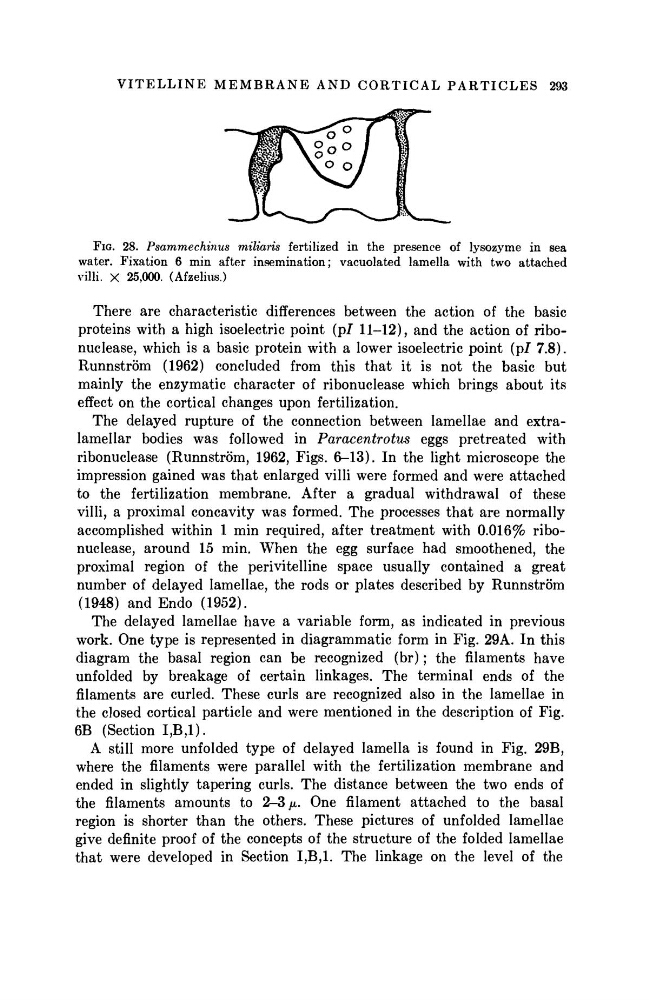
FIG. 28. Psammechinus miliaris fertilized in the presence of lysozyme in sea
water. Fixation 6 min after insemination; vacuolated lamella with two attached
villi. X 25,000. (Afzelius.)
There are characteristic differences between the action of the basic
proteins with a high isoelectric point (pi 11-12), and the action of ribonuclease, which is a basic protein with a lower isoelectric point (pi 7.8).
Runnström (1962) concluded from this that it is not the basic but
mainly the enzymatic character of ribonuclease which brings about its
effect on the cortical changes upon fertilization.
The delayed rupture of the connection between lamellae and extralamellar bodies was followed in Paracentrotus
eggs pretreated with
ribonuclease (Runnström, 1962, Figs. 6-13). In the light microscope the
impression gained was that enlarged villi were formed and were attached
to the fertilization membrane. After a gradual withdrawal of these
villi, a proximal concavity was formed. The processes that are normally
accomplished within 1 min required, after treatment with 0.016% ribonuclease, around 15 min. When the egg surface had smoothened, the
proximal region of the perivitelline space usually contained a great
number of delayed lamellae, the rods or plates described by Runnström
(1948) and Endo (1952).
The delayed lamellae have a variable form, as indicated in previous
work. One type is represented in diagrammatic form in Fig. 29A. In this
diagram the basal region can be recognized (br) ; the filaments have
unfolded by breakage of certain linkages. The terminal ends of the
filaments are curled. These curls are recognized also in the lamellae in
the closed cortical particle and were mentioned in the description of Fig.
6B (Section Ι,Β,Ι).
A still more unfolded type of delayed lamella is found in Fig. 29B,
where the filaments were parallel with the fertilization membrane and
ended in slightly tapering curls. The distance between the two ends of
the filaments amounts to 2-3 μ. One filament attached to the basal
region is shorter than the others. These pictures of unfolded lamellae
give definite proof of the concepts of the structure of the folded lamellae
that were developed in Section Ι,Β,Ι. The linkage on the level of the
FIG. 28. Psammechinus miliaris fertilized in the presence of lysozyme in sea
water. Fixation 6 min after insemination; vacuolated lamella with two attached
villi. X 25,000. (Afzelius.)
There are characteristic differences between the action of the basic
proteins with a high isoelectric point (pi 11-12), and the action of ribonuclease, which is a basic protein with a lower isoelectric point (pi 7.8).
Runnström (1962) concluded from this that it is not the basic but
mainly the enzymatic character of ribonuclease which brings about its
effect on the cortical changes upon fertilization.
The delayed rupture of the connection between lamellae and extralamellar bodies was followed in Paracentrotus
eggs pretreated with
ribonuclease (Runnström, 1962, Figs. 6-13). In the light microscope the
impression gained was that enlarged villi were formed and were attached
to the fertilization membrane. After a gradual withdrawal of these
villi, a proximal concavity was formed. The processes that are normally
accomplished within 1 min required, after treatment with 0.016% ribonuclease, around 15 min. When the egg surface had smoothened, the
proximal region of the perivitelline space usually contained a great
number of delayed lamellae, the rods or plates described by Runnström
(1948) and Endo (1952).
The delayed lamellae have a variable form, as indicated in previous
work. One type is represented in diagrammatic form in Fig. 29A. In this
diagram the basal region can be recognized (br) ; the filaments have
unfolded by breakage of certain linkages. The terminal ends of the
filaments are curled. These curls are recognized also in the lamellae in
the closed cortical particle and were mentioned in the description of Fig.
6B (Section Ι,Β,Ι).
A still more unfolded type of delayed lamella is found in Fig. 29B,
where the filaments were parallel with the fertilization membrane and
ended in slightly tapering curls. The distance between the two ends of
the filaments amounts to 2-3 μ. One filament attached to the basal
region is shorter than the others. These pictures of unfolded lamellae
give definite proof of the concepts of the structure of the folded lamellae
that were developed in Section Ι,Β,Ι. The linkage on the level of the