

252
JOHN RUNNSTRÖM
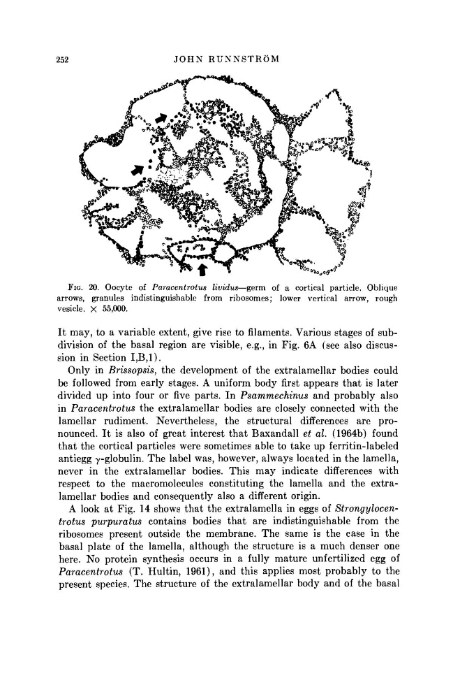
FIG. 20. Oocyte of Paracentrotus lividus—germ of a cortical particle. Oblique
arrows, granules indistinguishable from ribosomes; lower vertical arrow, rough
vesicle. X 55,000.
It may, to a variable extent, give rise to filaments. Various stages of subdivision of the basal region are visible, e.g., in Fig. 6A (see also discussion in Section Ι,Β,Ι).
Only in Brissopsis, the development of the extralamellar bodies could
be followed from early stages. A uniform body first appears that is later
divided up into four or five parts. In Psammechinus
and probably also
in Paracentrotus
the extralamellar bodies are closely connected with the
lamellar rudiment. Nevertheless, the structural differences are pronounced. It is also of great interest that Baxandall et al. (1964b) found
that the cortical particles were sometimes able to take up ferritin-labeled
antiegg γ-globulin. The label was, however, always located in the lamella,
never in the extralamellar bodies. This may indicate differences with
respect to the macromolecules constituting the lamella and the extralamellar bodies and consequently also a different origin.
A look at Fig. 14 shows that the extralamella in eggs of
Strongylocentrotus purpuratus
contains bodies that are indistinguishable from the
ribosomes present outside the membrane. The same is the case in the
basal plate of the lamella, although the structure is a much denser one
here. No protein synthesis occurs in a fully mature unfertilized egg of
Paracentrotus
(T. Hultin, 1961), and this applies most probably to the
present species. The structure of the extralamellar body and of the basal
JOHN RUNNSTRÖM
FIG. 20. Oocyte of Paracentrotus lividus—germ of a cortical particle. Oblique
arrows, granules indistinguishable from ribosomes; lower vertical arrow, rough
vesicle. X 55,000.
It may, to a variable extent, give rise to filaments. Various stages of subdivision of the basal region are visible, e.g., in Fig. 6A (see also discussion in Section Ι,Β,Ι).
Only in Brissopsis, the development of the extralamellar bodies could
be followed from early stages. A uniform body first appears that is later
divided up into four or five parts. In Psammechinus
and probably also
in Paracentrotus
the extralamellar bodies are closely connected with the
lamellar rudiment. Nevertheless, the structural differences are pronounced. It is also of great interest that Baxandall et al. (1964b) found
that the cortical particles were sometimes able to take up ferritin-labeled
antiegg γ-globulin. The label was, however, always located in the lamella,
never in the extralamellar bodies. This may indicate differences with
respect to the macromolecules constituting the lamella and the extralamellar bodies and consequently also a different origin.
A look at Fig. 14 shows that the extralamella in eggs of
Strongylocentrotus purpuratus
contains bodies that are indistinguishable from the
ribosomes present outside the membrane. The same is the case in the
basal plate of the lamella, although the structure is a much denser one
here. No protein synthesis occurs in a fully mature unfertilized egg of
Paracentrotus
(T. Hultin, 1961), and this applies most probably to the
present species. The structure of the extralamellar body and of the basal