

250
J O H N R U N N S T R Ö M
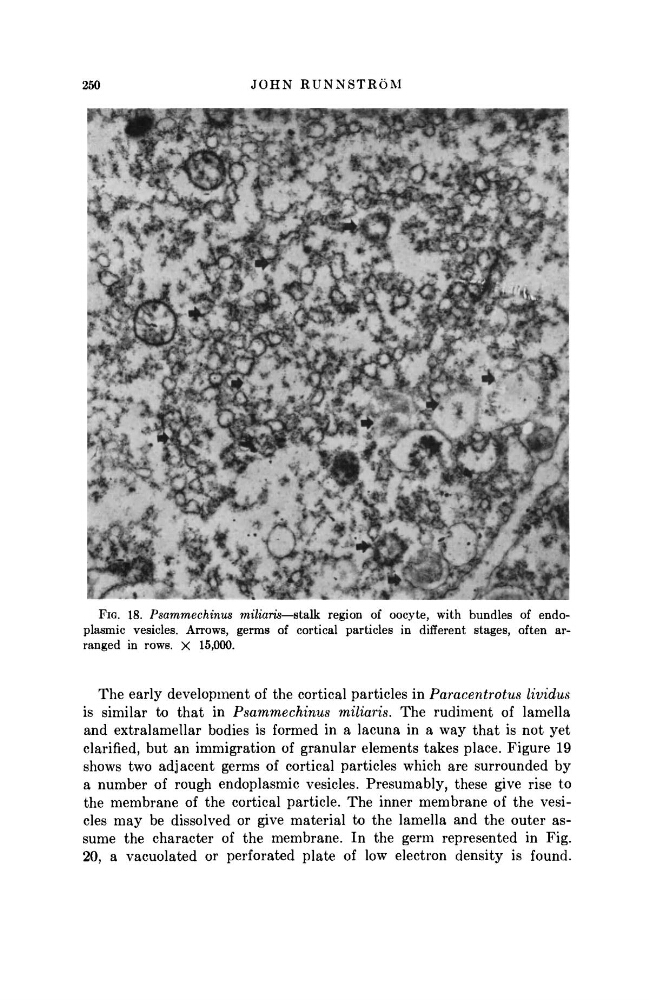
FIG. 18. Psammechinus miliaris—stalk region of oocyte, with bundles of endoplasmic vesicles. Arrows, germs of cortical particles in different stages, often arranged in rows. X 15,000.
The early development of the cortical particles in Paracentrotus
lividus
is similar to t h a t in Psammechinus
miliaris. The rudiment of lamella
and extralamellar bodies is formed in a lacuna in a way t h a t is not yet
clarified, but an immigration of granular elements takes place. Figure 19
shows two adjacent germs of cortical particles which are surrounded by
a number of rough endoplasmic vesicles. Presumably, these give rise to
the membrane of the cortical particle. The inner membrane of the vesicles may be dissolved or give material to the lamella and the outer assume the character of the membrane. In the germ represented in Fig.
20, a vacuolated or perforated plate of low electron density is found.
J O H N R U N N S T R Ö M
FIG. 18. Psammechinus miliaris—stalk region of oocyte, with bundles of endoplasmic vesicles. Arrows, germs of cortical particles in different stages, often arranged in rows. X 15,000.
The early development of the cortical particles in Paracentrotus
lividus
is similar to t h a t in Psammechinus
miliaris. The rudiment of lamella
and extralamellar bodies is formed in a lacuna in a way t h a t is not yet
clarified, but an immigration of granular elements takes place. Figure 19
shows two adjacent germs of cortical particles which are surrounded by
a number of rough endoplasmic vesicles. Presumably, these give rise to
the membrane of the cortical particle. The inner membrane of the vesicles may be dissolved or give material to the lamella and the outer assume the character of the membrane. In the germ represented in Fig.
20, a vacuolated or perforated plate of low electron density is found.