

V I T E L L I N E M E M B R A N E A N D C O R T I C A L P A R T I C L E S 249
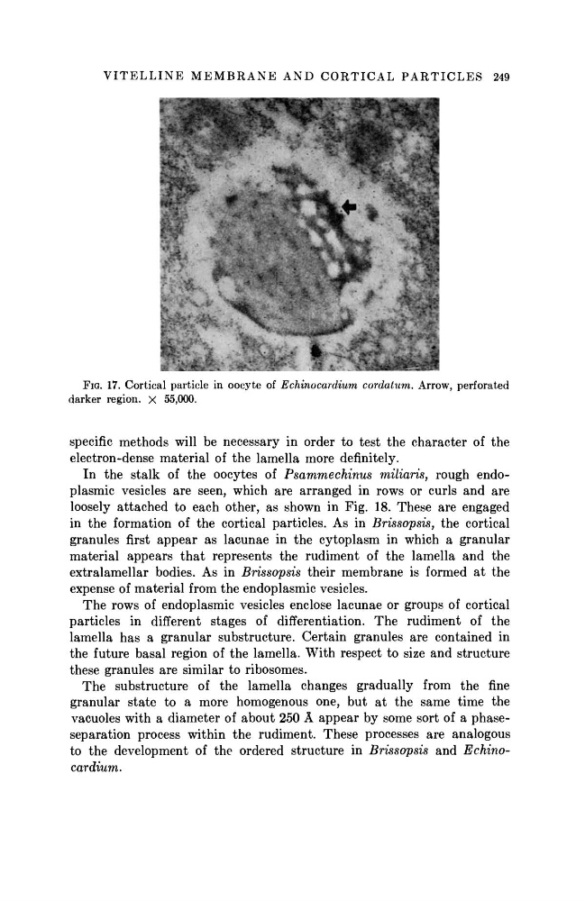
FIG. 17. Cortical particle in oocyte of Echinocardium cordatum. Arrow, perforated
darker region. X 55,000.
specific methods will be necessary in order to test the character of the
electron-dense material of the lamella more definitely.
In the stalk of the oocytes of Psammechinus
miliaris, rough endoplasmic vesicles are seen, which are arranged in rows or curls and are
loosely attached to each other, as shown in Fig. 18. These are engaged
in the formation of the cortical particles. As in Brissopsis, the cortical
granules first appear as lacunae in the cytoplasm in which a granular
material appears t h a t represents the rudiment of the lamella and the
extralamellar bodies. As in Brissopsis their membrane is formed at the
expense of material from the endoplasmic vesicles.
The rows of endoplasmic vesicles enclose lacunae or groups of cortical
particles in different stages of differentiation. The rudiment of the
lamella has a granular substructure. Certain granules are contained in
the future basal region of the lamella. With respect to size and structure
these granules are similar to ribosomes.
The substructure of the lamella changes gradually from the fine
granular state to a more homogenous one, but at the same time the
vacuoles with a diameter of about 250 Â appear by some sort of a phaseseparation process within the rudiment. These processes are analogous
to the development of the ordered structure in Brissopsis and Echinocardium.
FIG. 17. Cortical particle in oocyte of Echinocardium cordatum. Arrow, perforated
darker region. X 55,000.
specific methods will be necessary in order to test the character of the
electron-dense material of the lamella more definitely.
In the stalk of the oocytes of Psammechinus
miliaris, rough endoplasmic vesicles are seen, which are arranged in rows or curls and are
loosely attached to each other, as shown in Fig. 18. These are engaged
in the formation of the cortical particles. As in Brissopsis, the cortical
granules first appear as lacunae in the cytoplasm in which a granular
material appears t h a t represents the rudiment of the lamella and the
extralamellar bodies. As in Brissopsis their membrane is formed at the
expense of material from the endoplasmic vesicles.
The rows of endoplasmic vesicles enclose lacunae or groups of cortical
particles in different stages of differentiation. The rudiment of the
lamella has a granular substructure. Certain granules are contained in
the future basal region of the lamella. With respect to size and structure
these granules are similar to ribosomes.
The substructure of the lamella changes gradually from the fine
granular state to a more homogenous one, but at the same time the
vacuoles with a diameter of about 250 Â appear by some sort of a phaseseparation process within the rudiment. These processes are analogous
to the development of the ordered structure in Brissopsis and Echinocardium.