

248
JOHN RUNNSTRÖM
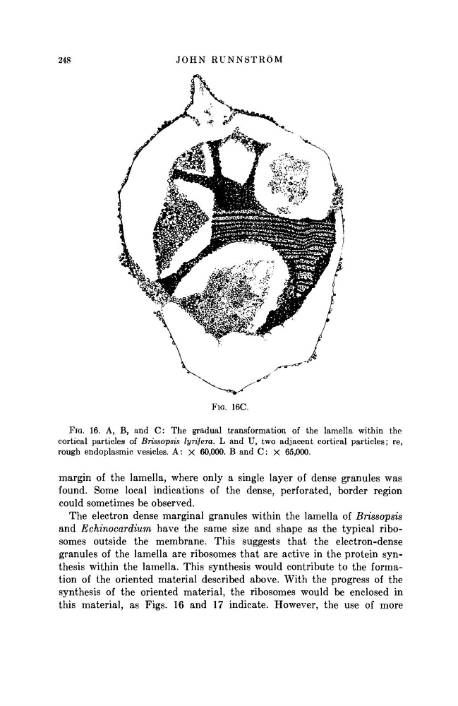
FIG. 16C.
FIG. 16. A, Β, and C: The gradual transformation of the lamella within the
cortical particles of Brissopsis lyrifera. L and U, two adjacent cortical particles; re,
rough endoplasmic vesicles. A: X 60,000. Β and C: χ 65,000.
margin of the lamella, where only a single layer of dense granules was
found. Some local indications of the dense, perforated, border region
could sometimes be observed.
The electron dense marginal granules within the lamella of Brissopsis
and Echinocardium
have the same size and shape as the typical ribosomes outside the membrane. This suggests that the electron-dense
granules of the lamella are ribosomes that are active in the protein synthesis within the lamella. This synthesis would contribute to the formation of the oriented material described above. With the progress of the
synthesis of the oriented material, the ribosomes would be enclosed in
this material, as Figs. 16 and 17 indicate. However, the use of more
JOHN RUNNSTRÖM
FIG. 16C.
FIG. 16. A, Β, and C: The gradual transformation of the lamella within the
cortical particles of Brissopsis lyrifera. L and U, two adjacent cortical particles; re,
rough endoplasmic vesicles. A: X 60,000. Β and C: χ 65,000.
margin of the lamella, where only a single layer of dense granules was
found. Some local indications of the dense, perforated, border region
could sometimes be observed.
The electron dense marginal granules within the lamella of Brissopsis
and Echinocardium
have the same size and shape as the typical ribosomes outside the membrane. This suggests that the electron-dense
granules of the lamella are ribosomes that are active in the protein synthesis within the lamella. This synthesis would contribute to the formation of the oriented material described above. With the progress of the
synthesis of the oriented material, the ribosomes would be enclosed in
this material, as Figs. 16 and 17 indicate. However, the use of more