

VITELLINE MEMBRANE AND CORTICAL PARTICLES
247
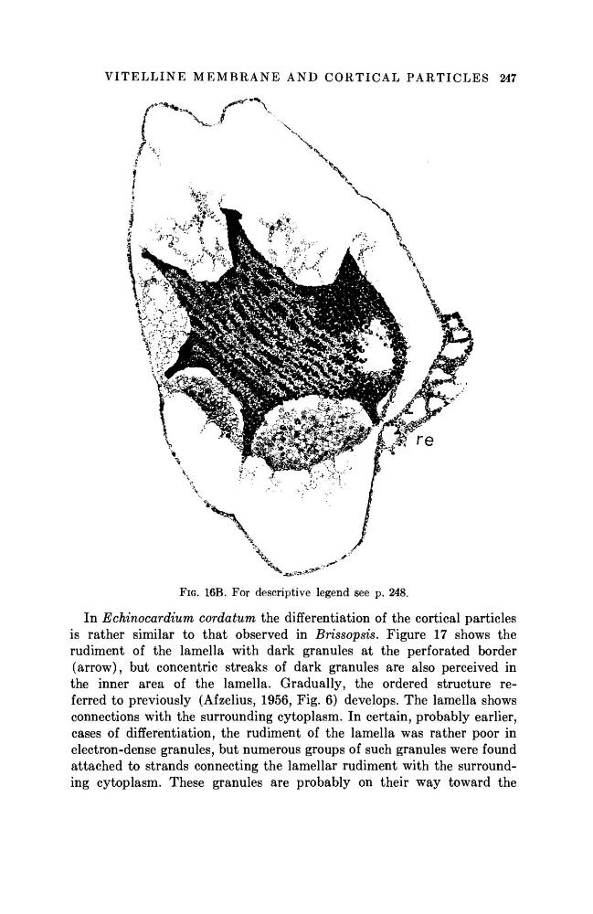
FIG. 16B. For descriptive legend see p. 248.
In Echinocardium
cordatum the differentiation of the cortical particles
is rather similar to that observed in Brissopsis. Figure 17 shows the
rudiment of the lamella with dark granules at the perforated border
(arrow), but concentric streaks of dark granules are also perceived in
the inner area of the lamella. Gradually, the ordered structure referred to previously (Afzelius, 1956, Fig. 6) develops. The lamella shows
connections with the surrounding cytoplasm. In certain, probably earlier,
cases of differentiation, the rudiment of the lamella was rather poor in
electron-dense granules, but numerous groups of such granules were found
attached to strands connecting the lamellar rudiment with the surrounding cytoplasm. These granules are probably on their way toward the
247
FIG. 16B. For descriptive legend see p. 248.
In Echinocardium
cordatum the differentiation of the cortical particles
is rather similar to that observed in Brissopsis. Figure 17 shows the
rudiment of the lamella with dark granules at the perforated border
(arrow), but concentric streaks of dark granules are also perceived in
the inner area of the lamella. Gradually, the ordered structure referred to previously (Afzelius, 1956, Fig. 6) develops. The lamella shows
connections with the surrounding cytoplasm. In certain, probably earlier,
cases of differentiation, the rudiment of the lamella was rather poor in
electron-dense granules, but numerous groups of such granules were found
attached to strands connecting the lamellar rudiment with the surrounding cytoplasm. These granules are probably on their way toward the