

246
JOHN RUNNSTRÖM
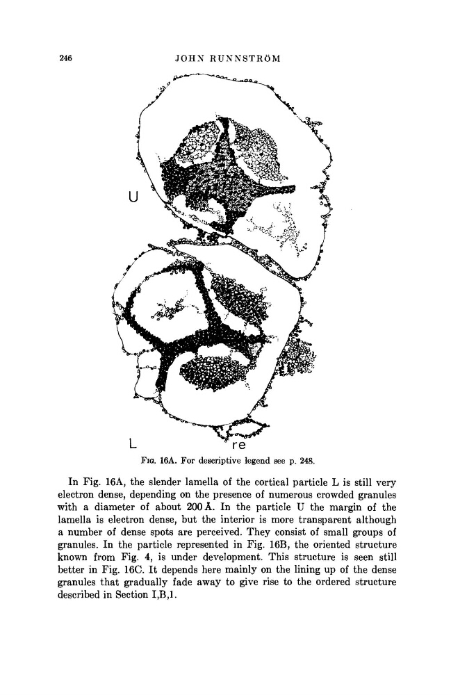
FIG. 16A. For descriptive legend see p. 248.
In Fig. 16A, the slender lamella of the cortical particle L is still very
electron dense, depending on the presence of numerous crowded granules
with a diameter of about 200 Â. In the particle U the margin of the
lamella is electron dense, but the interior is more transparent although
a number of dense spots are perceived. They consist of small groups of
granules. In the particle represented in Fig. 16B, the oriented structure
known from Fig. 4, is under development. This structure is seen still
better in Fig. 16C. It depends here mainly on the lining up of the dense
granules that gradually fade away to give rise to the ordered structure
described in Section Ι,Β,Ι.
JOHN RUNNSTRÖM
FIG. 16A. For descriptive legend see p. 248.
In Fig. 16A, the slender lamella of the cortical particle L is still very
electron dense, depending on the presence of numerous crowded granules
with a diameter of about 200 Â. In the particle U the margin of the
lamella is electron dense, but the interior is more transparent although
a number of dense spots are perceived. They consist of small groups of
granules. In the particle represented in Fig. 16B, the oriented structure
known from Fig. 4, is under development. This structure is seen still
better in Fig. 16C. It depends here mainly on the lining up of the dense
granules that gradually fade away to give rise to the ordered structure
described in Section Ι,Β,Ι.