

YOLK OF THE HEN'S EGG
239
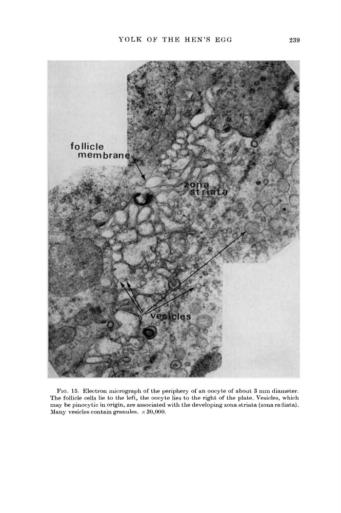
FIG. 15. Electron micrograph of the periphery of an oocyte of about 3 mm diameter.
The follicle cells lie to the left, the oocyte lies to the right of the plate. Vesicles, which
may be pinocytic in origin, are associated with the developing zona striata (zona radiata).
Many vesicles contain granules, x 30,000.
239
FIG. 15. Electron micrograph of the periphery of an oocyte of about 3 mm diameter.
The follicle cells lie to the left, the oocyte lies to the right of the plate. Vesicles, which
may be pinocytic in origin, are associated with the developing zona striata (zona radiata).
Many vesicles contain granules, x 30,000.