

238
RUTH BELLAIRS
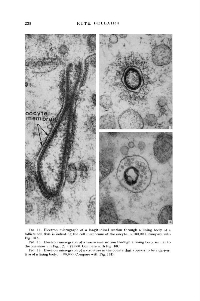
FIG. 12. Electron micrograph of a longitudinal section through a lining body of a
follicle cell that is indenting the cell membrane of the oocyte. x 130,000. Compare with
Fig. 16A.
FIG. 13. Electron micrograph of a transverse section through a lining body similar to
the one shown in Fig. 12. χ 73,000. Compare with Fig. 16C.
FIG. 14. Electron micrograph of a structure in the oocyte that appears to be a derivative of a lining body, x 80,000. Compare with Fig. 16D.
RUTH BELLAIRS
FIG. 12. Electron micrograph of a longitudinal section through a lining body of a
follicle cell that is indenting the cell membrane of the oocyte. x 130,000. Compare with
Fig. 16A.
FIG. 13. Electron micrograph of a transverse section through a lining body similar to
the one shown in Fig. 12. χ 73,000. Compare with Fig. 16C.
FIG. 14. Electron micrograph of a structure in the oocyte that appears to be a derivative of a lining body, x 80,000. Compare with Fig. 16D.