

16S
HENRIETTE HERLANT-MEEWIS
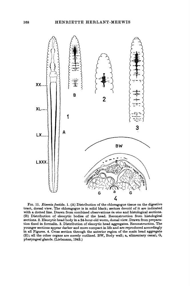
FIG. 11. Eisenia foetida. 1. (A) Distribution of the chloragogue tissue on the digestive
tract, dorsal view. The chloragogue is in solid black; sectors devoid of it are indicated
with a dotted line. Drawn from combined observations in vivo and histological sections.
(B) Distribution of eleocytic bodies of the head. Reconstruction from histological
sections. 2. Eleocytic head body in a 24-hour-old worm, dorsal view. Drawn from preparation fixed in formalin. 3. Distribution of eleocytic head aggregates. Reconstruction. The
younger sections appear darker and more compact in life and are reproduced accordingly
in all Figures. 4. Cross section through the anterior region of the main head aggregate
(E); all the other organs are merely outlined. BW, Body wall; a, alimentary canal; G,
pharyngeal glands. (Liebmann, 1943.)
HENRIETTE HERLANT-MEEWIS
FIG. 11. Eisenia foetida. 1. (A) Distribution of the chloragogue tissue on the digestive
tract, dorsal view. The chloragogue is in solid black; sectors devoid of it are indicated
with a dotted line. Drawn from combined observations in vivo and histological sections.
(B) Distribution of eleocytic bodies of the head. Reconstruction from histological
sections. 2. Eleocytic head body in a 24-hour-old worm, dorsal view. Drawn from preparation fixed in formalin. 3. Distribution of eleocytic head aggregates. Reconstruction. The
younger sections appear darker and more compact in life and are reproduced accordingly
in all Figures. 4. Cross section through the anterior region of the main head aggregate
(E); all the other organs are merely outlined. BW, Body wall; a, alimentary canal; G,
pharyngeal glands. (Liebmann, 1943.)