

R E G E N E R A T I O N I N A N N E L I D S
167
incapable of dividing and consequently there is no expansion of the
walls. After 4 weeks, amoebocytes that have migrated from nonirradiated anterior segments penetrate the regeneration bud in succession and contribute to its normal growth. Stephan-Dubois concluded
from these observations that some free cells in the coelom can play the
same role as neoblasts ; these totipotent amoebocytes could participate
in the reconstruction of all organs in the regeneration bud.
This concept had already been proposed by Dehorne (1949). Recently,
Clark and Clark (1962) have also described the change of coelomocytes
into fibroblasts. However, these would participate in mesodermal
regeneration only. The amoebocytes described by Stephan-Dubois are
not the only mesodermal cells to have attracted the attention of workers
studying regeneration phenomena. Since Nusbaum's observations
(1908), several investigators have described the accumulation of various
coelomic elements near the wound-healing zone (Romieu, 1923; FauréFremiet, 1927; Ohuye, 1937-38). According to Fauré-Fremiet, some
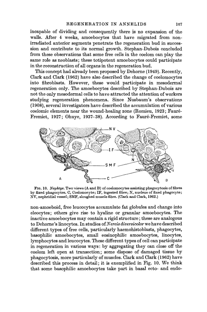
FIG. 10. Nephtys. Two views (A and B) of coelomocytes assisting phagocytosis of fibres
by fixed phagocytes. C, Coelomocy te ; IF, ingested fibre; N, nucleus of fixed phagocyte;
NV, nephridial vessel; SMF, sloughed muscle fibre. (Clark and Clark, 1962.)
non-amoeboid, free leucocytes accumulate fat globules and change into
eleocytes; others give rise to hyaline or granular amoebocytes. The
inactive amoebocytes may contain a rigid structure ; these are analogous
to Dehorne's linocytes. In studies of Nereis diversicolor we have described
different types of free cells, particularly haemohistoblasts, phagocytes,
basophilic amoebocytes, small eosinophilic amoebocytes, linocytes,
lymphocytes and leucocytes. These different types of cell can participate
in regeneration in various ways : by aggregating they can close off the
coelom left open at transection; some dispose of damaged tissue by
phagocytosis, more particularly of muscles. Clark and Clark (1962) have
described this process in detail; it is exemplified in Fig. 10. We think
that some basophilic amoebocytes take part in basal ecto- and endo-
167
incapable of dividing and consequently there is no expansion of the
walls. After 4 weeks, amoebocytes that have migrated from nonirradiated anterior segments penetrate the regeneration bud in succession and contribute to its normal growth. Stephan-Dubois concluded
from these observations that some free cells in the coelom can play the
same role as neoblasts ; these totipotent amoebocytes could participate
in the reconstruction of all organs in the regeneration bud.
This concept had already been proposed by Dehorne (1949). Recently,
Clark and Clark (1962) have also described the change of coelomocytes
into fibroblasts. However, these would participate in mesodermal
regeneration only. The amoebocytes described by Stephan-Dubois are
not the only mesodermal cells to have attracted the attention of workers
studying regeneration phenomena. Since Nusbaum's observations
(1908), several investigators have described the accumulation of various
coelomic elements near the wound-healing zone (Romieu, 1923; FauréFremiet, 1927; Ohuye, 1937-38). According to Fauré-Fremiet, some
FIG. 10. Nephtys. Two views (A and B) of coelomocytes assisting phagocytosis of fibres
by fixed phagocytes. C, Coelomocy te ; IF, ingested fibre; N, nucleus of fixed phagocyte;
NV, nephridial vessel; SMF, sloughed muscle fibre. (Clark and Clark, 1962.)
non-amoeboid, free leucocytes accumulate fat globules and change into
eleocytes; others give rise to hyaline or granular amoebocytes. The
inactive amoebocytes may contain a rigid structure ; these are analogous
to Dehorne's linocytes. In studies of Nereis diversicolor we have described
different types of free cells, particularly haemohistoblasts, phagocytes,
basophilic amoebocytes, small eosinophilic amoebocytes, linocytes,
lymphocytes and leucocytes. These different types of cell can participate
in regeneration in various ways : by aggregating they can close off the
coelom left open at transection; some dispose of damaged tissue by
phagocytosis, more particularly of muscles. Clark and Clark (1962) have
described this process in detail; it is exemplified in Fig. 10. We think
that some basophilic amoebocytes take part in basal ecto- and endo-