

R E G E N E R A T I O N I N A N N E L I D S
169
dermal cell destruction. This could also be the role of the coelomocytes
shown by Clark and Clark in the cicatricial ectoderm.
Liebmann (1942a, b, 1943, 1946) has paid special attention to cells
which he called eleocytes or trephocytes. He contrasted them with
lymphoidocytes. According to Liebmann, eleocytes are derived from
chloragogue tissue where nutritious substances are elaborated. Loaded
with storage products, the eleocytes are set free and carry these substances to all organs, thus having an important trophic function. In the
cephalic region, they form polarized aggregates whose arrangement is
shown in Fig. 11. The younger cells, newly released, gather towards the
most anterior region while posteriorly the aggregate breaks up to give
rise to free eleocytes. These aggregates consist essentially of eleocytes
together with some amoebocytes. They already exist in the worm when
it emerges from the cocoon. Although not clearly delimited, they
occupy a well-defined metameric position. Their appearance and position can be seen in Fig. 11(2). When a worm is transected in the cephalic
region, the aggregate undergoes a series of transformations. It breaks up,
and the cells which gather in the healing zone release some of their
storage granules while some undergo a sort of disintegration and are
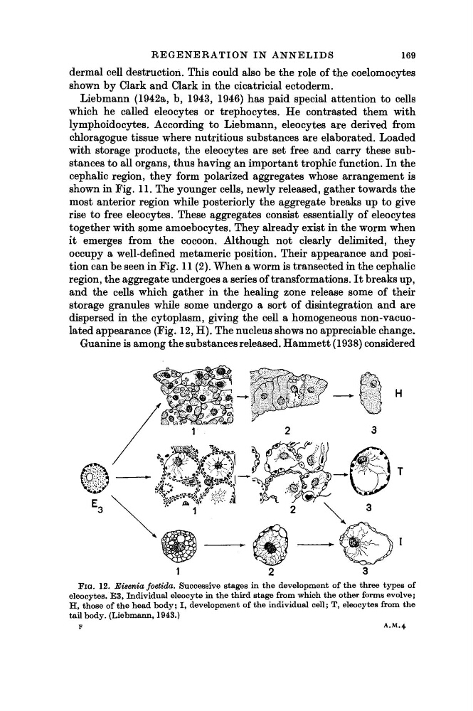
dispersed in the cytoplasm, giving the cell a homogeneous non-vacuolated appearance (Fig. 12, H). The nucleus shows no appreciable change.
Guanine is among the substances released. Hammett (1938) considered
FIG. 12. Eisenia foetida. Successive stages in the development of the three types of
eleocytes. E3, Individual eleocyte in the third stage from which the other forms evolve;
H, those of the head body; I, development of the individual cell; T, eleocytes from the
tail body. (Liebmann, 1943.)
F
A.M.4
169
dermal cell destruction. This could also be the role of the coelomocytes
shown by Clark and Clark in the cicatricial ectoderm.
Liebmann (1942a, b, 1943, 1946) has paid special attention to cells
which he called eleocytes or trephocytes. He contrasted them with
lymphoidocytes. According to Liebmann, eleocytes are derived from
chloragogue tissue where nutritious substances are elaborated. Loaded
with storage products, the eleocytes are set free and carry these substances to all organs, thus having an important trophic function. In the
cephalic region, they form polarized aggregates whose arrangement is
shown in Fig. 11. The younger cells, newly released, gather towards the
most anterior region while posteriorly the aggregate breaks up to give
rise to free eleocytes. These aggregates consist essentially of eleocytes
together with some amoebocytes. They already exist in the worm when
it emerges from the cocoon. Although not clearly delimited, they
occupy a well-defined metameric position. Their appearance and position can be seen in Fig. 11(2). When a worm is transected in the cephalic
region, the aggregate undergoes a series of transformations. It breaks up,
and the cells which gather in the healing zone release some of their
storage granules while some undergo a sort of disintegration and are
dispersed in the cytoplasm, giving the cell a homogeneous non-vacuolated appearance (Fig. 12, H). The nucleus shows no appreciable change.
Guanine is among the substances released. Hammett (1938) considered
FIG. 12. Eisenia foetida. Successive stages in the development of the three types of
eleocytes. E3, Individual eleocyte in the third stage from which the other forms evolve;
H, those of the head body; I, development of the individual cell; T, eleocytes from the
tail body. (Liebmann, 1943.)
F
A.M.4