

160
H E N R I E T T E HERLANT-MEEWIS
number of epithelial cells are subjected to histolysis. Some amoebocytes
enter the basal region of the walls and may participate in this breakdown by secreting lytic enzymes. Clark and Clark (1962) have also
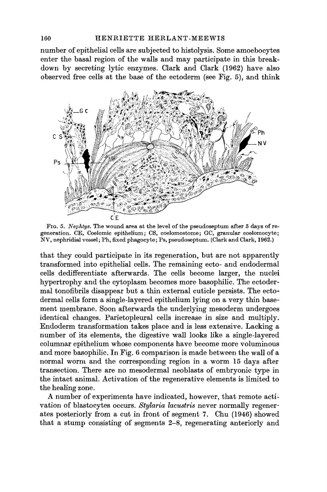
observed free cells at the base of the ectoderm (see Fig. 5), and think
F I G . 5. Nephtys. The wound area at the level of the pseudoseptum after 5 days of regeneration. CE, Coelomic epithelium; CS, coelomostome; GC, granular coelomocyte;
NV, nephridial vessel; Ph, fixed phagocyte; Ps, pseudoseptum. (Clark and Clark, 1962.)
that they could participate in its regeneration, but are not apparently
transformed into epithelial cells. The remaining ecto- and endodermal
cells dedifferentiate afterwards. The cells become larger, the nuclei
hypertrophy and the cytoplasm becomes more basophilic. The ectodermal tonofibrils disappear but a thin external cuticle persists. The ectodermal cells form a single-layered epithelium lying on a very thin basement membrane. Soon afterwards the underlying mesoderm undergoes
identical changes. Parietopleural cells increase in size and multiply.
Endoderm transformation takes place and is less extensive. Lacking a
number of its elements, the digestive wall looks like a single-layered
columnar epithelium whose components have become more voluminous
and more basophilic. In Fig. 6 comparison is made between the wall of a
normal worm and the corresponding region in a worm 15 days after
transection. There are no mesodermal neoblasts of embryonic type in
the intact animal. Activation of the regenerative elements is limited to
the healing zone.
A number of experiments have indicated, however, that remote activation of blastocytes occurs. Stylaria lacustris never normally regenerates posteriorly from a cut in front of segment 7. Chu (1946) showed
that a stump consisting of segments 2-8, regenerating anteriorly and
H E N R I E T T E HERLANT-MEEWIS
number of epithelial cells are subjected to histolysis. Some amoebocytes
enter the basal region of the walls and may participate in this breakdown by secreting lytic enzymes. Clark and Clark (1962) have also
observed free cells at the base of the ectoderm (see Fig. 5), and think
F I G . 5. Nephtys. The wound area at the level of the pseudoseptum after 5 days of regeneration. CE, Coelomic epithelium; CS, coelomostome; GC, granular coelomocyte;
NV, nephridial vessel; Ph, fixed phagocyte; Ps, pseudoseptum. (Clark and Clark, 1962.)
that they could participate in its regeneration, but are not apparently
transformed into epithelial cells. The remaining ecto- and endodermal
cells dedifferentiate afterwards. The cells become larger, the nuclei
hypertrophy and the cytoplasm becomes more basophilic. The ectodermal tonofibrils disappear but a thin external cuticle persists. The ectodermal cells form a single-layered epithelium lying on a very thin basement membrane. Soon afterwards the underlying mesoderm undergoes
identical changes. Parietopleural cells increase in size and multiply.
Endoderm transformation takes place and is less extensive. Lacking a
number of its elements, the digestive wall looks like a single-layered
columnar epithelium whose components have become more voluminous
and more basophilic. In Fig. 6 comparison is made between the wall of a
normal worm and the corresponding region in a worm 15 days after
transection. There are no mesodermal neoblasts of embryonic type in
the intact animal. Activation of the regenerative elements is limited to
the healing zone.
A number of experiments have indicated, however, that remote activation of blastocytes occurs. Stylaria lacustris never normally regenerates posteriorly from a cut in front of segment 7. Chu (1946) showed
that a stump consisting of segments 2-8, regenerating anteriorly and