

REGENERATION IN ANNELIDS
161
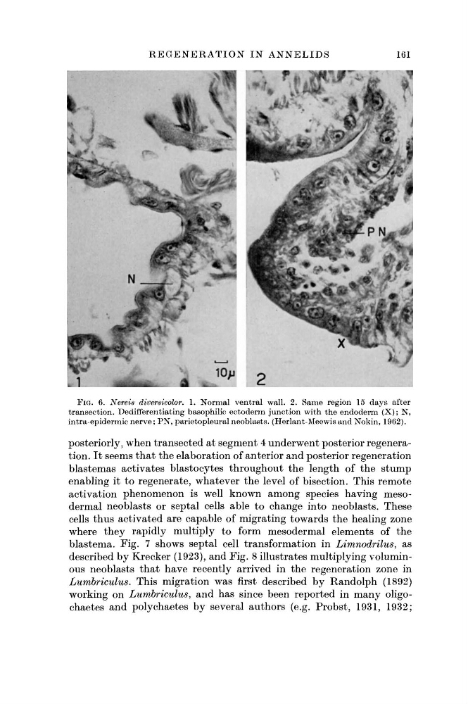
FIG. 6. Nereis diversicolor. 1. Normal ventral wall. 2. Same region 15 days after
transection. Dedifferentiating basophilic ectoderm junction with the endoderm (X); N,
intra-epidermic nerve; PN, parietopleural neoblasts. (Herlant-Meewis and Nokin, 1962).
posteriorly, when transected at segment 4 underwent posterior regeneration. It seems that the elaboration of anterior and posterior regeneration
blastemas activates blastocytes throughout the length of the stump
enabling it to regenerate, whatever the level of bisection. This remote
activation phenomenon is well known among species having mesodermal neoblasts or septal cells able to change into neoblasts. These
cells thus activated are capable of migrating towards the healing zone
where they rapidly multiply to form mesodermal elements of the
blastema. Fig. 7 shows septal cell transformation in Limnodrilus, as
described by Krecker (1923), and Fig. 8 illustrates multiplying voluminous neoblasts that have recently arrived in the regeneration zone in
Lumbriculus. This migration was first described by Randolph (1892)
working on Lumbriculus, and has since been reported in many oligochaetes and polychaetes by several authors (e.g. Probst, 1931, 1932;
161
FIG. 6. Nereis diversicolor. 1. Normal ventral wall. 2. Same region 15 days after
transection. Dedifferentiating basophilic ectoderm junction with the endoderm (X); N,
intra-epidermic nerve; PN, parietopleural neoblasts. (Herlant-Meewis and Nokin, 1962).
posteriorly, when transected at segment 4 underwent posterior regeneration. It seems that the elaboration of anterior and posterior regeneration
blastemas activates blastocytes throughout the length of the stump
enabling it to regenerate, whatever the level of bisection. This remote
activation phenomenon is well known among species having mesodermal neoblasts or septal cells able to change into neoblasts. These
cells thus activated are capable of migrating towards the healing zone
where they rapidly multiply to form mesodermal elements of the
blastema. Fig. 7 shows septal cell transformation in Limnodrilus, as
described by Krecker (1923), and Fig. 8 illustrates multiplying voluminous neoblasts that have recently arrived in the regeneration zone in
Lumbriculus. This migration was first described by Randolph (1892)
working on Lumbriculus, and has since been reported in many oligochaetes and polychaetes by several authors (e.g. Probst, 1931, 1932;