

REGENERATION IN ANNELIDS
159
outside a budding zone. Ectodermal cells slide over the wound and close
it. At the same time ordinary cells of the ectoderm, as well as those that
have begun to change into glandular or pigment cells, begin to dedifferentiate. The changes start on the ventral side near the nerve cord in the
skin and gradually advance towards the dorsal side. Endodermal cells
undergo the same changes simultaneously: the appearance of these
walls 9 h after transection is illustrated in Fig. 2. The mesoderm does
not become activated until 48 h after injury (Fig. 3). The entire blastema is then made up of identical elements, regardless of their origin.
Tissues of Nereis diversicolor do not contain replacement cells : regenerative components of the blastema are derived exclusively from epithelial
cell dedifferentiation. By exposing the animals to low temperature
(8-10° C) in order to slow down the healing processes we have been able
to analyse regenerative phenomena more easily.
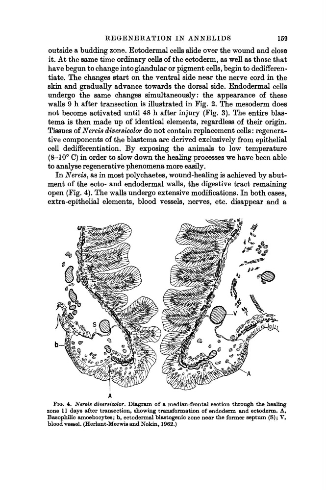
In Nereis, as in most polychaetes, wound-healing is achieved by abutment of the ecto- and endodermal walls, the digestive tract remaining
open (Fig. 4). The walls undergo extensive modifications. In both cases,
extra-epithelial elements, blood vessels, nerves, etc. disappear and a
FIG. 4. Nereis divereicolor. Diagram of a median-frontal section through the healing
zone 11 days after transection, showing transformation of endoderm and ectoderm. A,
Basophilic amoebocytes; b, ectodermal blastogenic zone near the former septum (S); V,
bloodvessel. (Herlant-MeewisandNokin, 1962.)
159
outside a budding zone. Ectodermal cells slide over the wound and close
it. At the same time ordinary cells of the ectoderm, as well as those that
have begun to change into glandular or pigment cells, begin to dedifferentiate. The changes start on the ventral side near the nerve cord in the
skin and gradually advance towards the dorsal side. Endodermal cells
undergo the same changes simultaneously: the appearance of these
walls 9 h after transection is illustrated in Fig. 2. The mesoderm does
not become activated until 48 h after injury (Fig. 3). The entire blastema is then made up of identical elements, regardless of their origin.
Tissues of Nereis diversicolor do not contain replacement cells : regenerative components of the blastema are derived exclusively from epithelial
cell dedifferentiation. By exposing the animals to low temperature
(8-10° C) in order to slow down the healing processes we have been able
to analyse regenerative phenomena more easily.
In Nereis, as in most polychaetes, wound-healing is achieved by abutment of the ecto- and endodermal walls, the digestive tract remaining
open (Fig. 4). The walls undergo extensive modifications. In both cases,
extra-epithelial elements, blood vessels, nerves, etc. disappear and a
FIG. 4. Nereis divereicolor. Diagram of a median-frontal section through the healing
zone 11 days after transection, showing transformation of endoderm and ectoderm. A,
Basophilic amoebocytes; b, ectodermal blastogenic zone near the former septum (S); V,
bloodvessel. (Herlant-MeewisandNokin, 1962.)