

352
Z. RYCHTER
the dorsal wall of the descending aorta, while a pair of intercostal
arteries branch from the rest of the evanescent left dorsal aorta; this
means that the beginning of the primitive subclavians in Fig. 21b is
most probably a remnant of the evanescent right dorsal aorta. For this
reason the mechanical torsion of the cranial part of the descending aorta
is not considered to be the cause of the shift. In our opinion, the
substitute arcus aortae may change the haemodynamic situation, which
may then cause the shift either directly or indirectly by a differential
growth of the aortic wall.
2. Carotid Artery as a Branch of the Pulmonary Arch
There are two types of this experimentally shifted origin of the
carotid arteries (see also Stephan, 1952). The first, regularly occurring
after suppression of both the 3rd and 4th aortic arches on either side,
is characterized by the dorsal position of the carotid root as a prolongation, in a cranial direction, of the dorsal aorta (e.g., Fig. 10, Fig. 15—
4 D
3 D
3 S
4
S ) . The second (already mentioned in Section II, B) occurred
once in 4-6 embryos in those combinations where all aortic arches of
one side were suppressed: the carotid artery is prolonged by the ventral
part of the corresponding 6th aortic arch, the pulmonary artery being
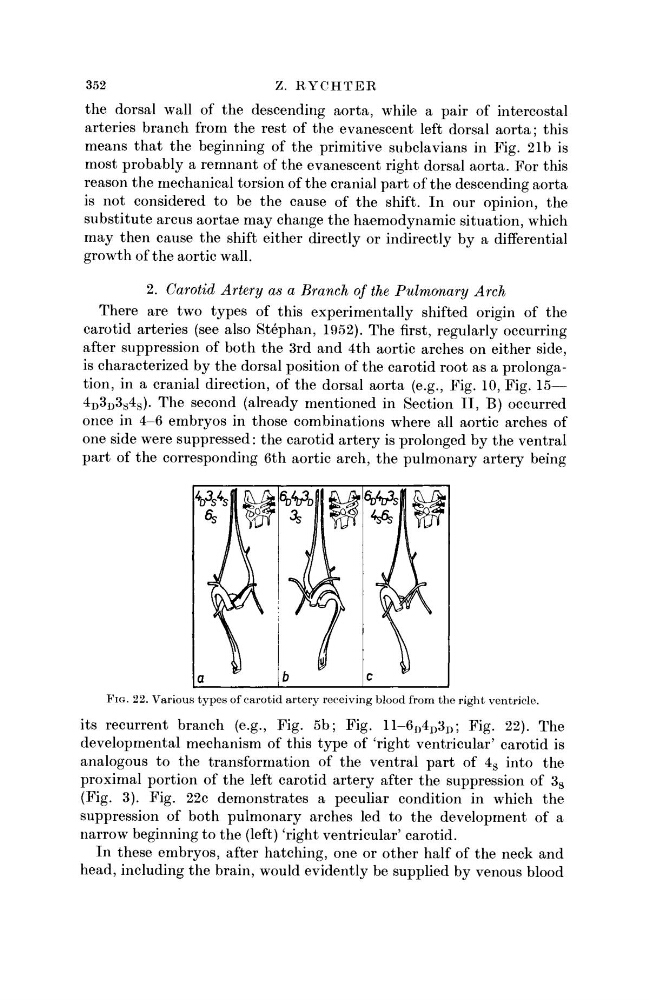
FIG. 22. Various types of carotid artery receiving blood from the right ventricle.
its recurrent branch (e.g., Fig. 5b; Fig. 11-6 D 4 D 3 D ; Fig. 22). The
developmental mechanism of this type of 'right ventricular' carotid is
analogous to the transformation of the ventral part of 4 S into the
proximal portion of the left carotid artery after the suppression of 3 S
(Fig. 3). Fig. 22c demonstrates a peculiar condition in which the
suppression of both pulmonary arches led to the development of a
narrow beginning to the (left) 'right ventricular' carotid.
In these embryos, after hatching, one or other half of the neck and
head, including the brain, would evidently be supplied by venous blood
Z. RYCHTER
the dorsal wall of the descending aorta, while a pair of intercostal
arteries branch from the rest of the evanescent left dorsal aorta; this
means that the beginning of the primitive subclavians in Fig. 21b is
most probably a remnant of the evanescent right dorsal aorta. For this
reason the mechanical torsion of the cranial part of the descending aorta
is not considered to be the cause of the shift. In our opinion, the
substitute arcus aortae may change the haemodynamic situation, which
may then cause the shift either directly or indirectly by a differential
growth of the aortic wall.
2. Carotid Artery as a Branch of the Pulmonary Arch
There are two types of this experimentally shifted origin of the
carotid arteries (see also Stephan, 1952). The first, regularly occurring
after suppression of both the 3rd and 4th aortic arches on either side,
is characterized by the dorsal position of the carotid root as a prolongation, in a cranial direction, of the dorsal aorta (e.g., Fig. 10, Fig. 15—
4 D
3 D
3 S
4
S ) . The second (already mentioned in Section II, B) occurred
once in 4-6 embryos in those combinations where all aortic arches of
one side were suppressed: the carotid artery is prolonged by the ventral
part of the corresponding 6th aortic arch, the pulmonary artery being
FIG. 22. Various types of carotid artery receiving blood from the right ventricle.
its recurrent branch (e.g., Fig. 5b; Fig. 11-6 D 4 D 3 D ; Fig. 22). The
developmental mechanism of this type of 'right ventricular' carotid is
analogous to the transformation of the ventral part of 4 S into the
proximal portion of the left carotid artery after the suppression of 3 S
(Fig. 3). Fig. 22c demonstrates a peculiar condition in which the
suppression of both pulmonary arches led to the development of a
narrow beginning to the (left) 'right ventricular' carotid.
In these embryos, after hatching, one or other half of the neck and
head, including the brain, would evidently be supplied by venous blood