

E X P E R I M E N T S ON EMBRYONIC AORTIC ARCHES 353
from the right ventricle, particularly since no ventricular defect was
observed in the majority of the combinations.
3. Relation of the Primitive and Definitive Subclavian Arteries
Information on the development of subclavian arteries in birds is
given in the publications of Hochstetter (1890), Bakst and Chafee
(1928) and Fleming (1926). Experimentally evoked anomalies of the
subclavian arteries were first described by Stephan (1952). Rychter and
Lemez (1962b) had 812 experimental embryos at their disposal for the
study of the occurrence and morphology of anomalous subclavians.
From the large number of different subclavian artery anomalies only
the relations between the primitive and definitive subclavian arteries
are reported here, for their haemodynamic significance.
Persistence of the primitive subclavian arteries only, with failure of
the definite subclavians to develop was observed in 92 cases (i.e., persistence occurred in 11-3%), bilaterally in 42 embryos, on the left in 33
embryos, and on the right in 17.
The persistence of a primitive subclavian artery was related neither
to the suppression of the 3rd aortic arches (from which the definitive
subclavian originates) nor to the side of experimental intervention.
The number of suppressed aortic arches, however, was directly related
to the frequency of primitive subclavian persistence. The persistence,
therefore, is most probably caused haemodynamically. Nevertheless,
no correlation with the incidence of ventricular defects was ascertained.
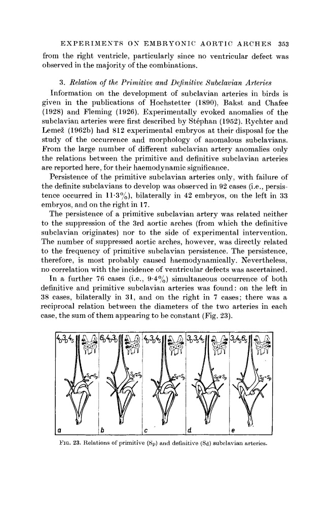
In a further 76 cases (i.e., 9-4%) simultaneous occurrence of both
definitive and primitive subclavian arteries was found: on the left in
38 cases, bilaterally in 31, and on the right in 7 cases; there was a
reciprocal relation between the diameters of the two arteries in each
case, the sum of them appearing to be constant (Fig. 23).
FIG. 23. Relations of primitive (Sp) and definitive (Sd) subclavian arteries.
from the right ventricle, particularly since no ventricular defect was
observed in the majority of the combinations.
3. Relation of the Primitive and Definitive Subclavian Arteries
Information on the development of subclavian arteries in birds is
given in the publications of Hochstetter (1890), Bakst and Chafee
(1928) and Fleming (1926). Experimentally evoked anomalies of the
subclavian arteries were first described by Stephan (1952). Rychter and
Lemez (1962b) had 812 experimental embryos at their disposal for the
study of the occurrence and morphology of anomalous subclavians.
From the large number of different subclavian artery anomalies only
the relations between the primitive and definitive subclavian arteries
are reported here, for their haemodynamic significance.
Persistence of the primitive subclavian arteries only, with failure of
the definite subclavians to develop was observed in 92 cases (i.e., persistence occurred in 11-3%), bilaterally in 42 embryos, on the left in 33
embryos, and on the right in 17.
The persistence of a primitive subclavian artery was related neither
to the suppression of the 3rd aortic arches (from which the definitive
subclavian originates) nor to the side of experimental intervention.
The number of suppressed aortic arches, however, was directly related
to the frequency of primitive subclavian persistence. The persistence,
therefore, is most probably caused haemodynamically. Nevertheless,
no correlation with the incidence of ventricular defects was ascertained.
In a further 76 cases (i.e., 9-4%) simultaneous occurrence of both
definitive and primitive subclavian arteries was found: on the left in
38 cases, bilaterally in 31, and on the right in 7 cases; there was a
reciprocal relation between the diameters of the two arteries in each
case, the sum of them appearing to be constant (Fig. 23).
FIG. 23. Relations of primitive (Sp) and definitive (Sd) subclavian arteries.