

E X P E R I M E N T S ON E M B R Y O N I C A O R T I C A R C H E S 351
nearer to the midline (Fig. 19b). In the combination 6 D 4 D 3 D 3 S (Fig.
19a, b) it is evident that the retention of the original position in the midline is dependent on the completeness of the arcus aortae from 4s.
Microdissection of the coeliac plexus in some normal and experimental embryos showed, in contradiction to the opinion of Bremer
(1926), that the sympathetic nerve fibres follow passively the changing
position of the coeliac root (Fig. 20).
FIG. 20. The relation of sympathetic nerve fibres of the coeliac plexus to the coeliac
root in various positions.
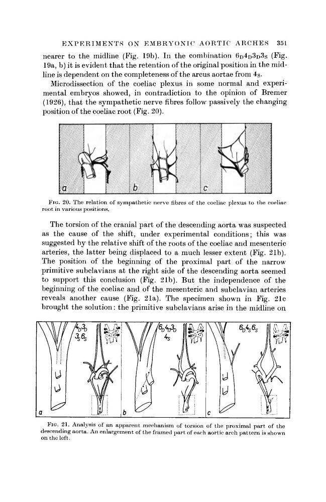
The torsion of the cranial part of the descending aorta was suspected
as the cause of the shift, under experimental conditions; this was
suggested by the relative shift of the roots of the coeliac and mesenteric
arteries, the latter being displaced to a much lesser extent (Fig. 21b).
The position of the beginning of the proximal part of the narrow
primitive subclavians at the right side of the descending aorta seemed
to support this conclusion (Fig. 21b). But the independence of the
beginning of the coeliac and of the mesenteric and subclavian arteries
reveals another cause (Fig. 21a). The specimen shown in Fig. 21c
brought the solution: the primitive subclavians arise in the midline on
FIG. 21. Analysis of an apparent mechanism of torsion of the proximal part of the
descending aorta. An enlargement of the framed part of each aortic arch pattern is shown
on the left.
nearer to the midline (Fig. 19b). In the combination 6 D 4 D 3 D 3 S (Fig.
19a, b) it is evident that the retention of the original position in the midline is dependent on the completeness of the arcus aortae from 4s.
Microdissection of the coeliac plexus in some normal and experimental embryos showed, in contradiction to the opinion of Bremer
(1926), that the sympathetic nerve fibres follow passively the changing
position of the coeliac root (Fig. 20).
FIG. 20. The relation of sympathetic nerve fibres of the coeliac plexus to the coeliac
root in various positions.
The torsion of the cranial part of the descending aorta was suspected
as the cause of the shift, under experimental conditions; this was
suggested by the relative shift of the roots of the coeliac and mesenteric
arteries, the latter being displaced to a much lesser extent (Fig. 21b).
The position of the beginning of the proximal part of the narrow
primitive subclavians at the right side of the descending aorta seemed
to support this conclusion (Fig. 21b). But the independence of the
beginning of the coeliac and of the mesenteric and subclavian arteries
reveals another cause (Fig. 21a). The specimen shown in Fig. 21c
brought the solution: the primitive subclavians arise in the midline on
FIG. 21. Analysis of an apparent mechanism of torsion of the proximal part of the
descending aorta. An enlargement of the framed part of each aortic arch pattern is shown
on the left.