

350
Z. RYCHTER
It is apparent (Fig. 17—compare 6 D 4 D and 6 D 4 D 3 D ) that the root of
the coeliac artery remained in its early original midline position when
no arcus aortae (including 6 D ) developed on the right side and when the
substitute arcus aortae was formed by 4 S alone (Fig. 17b). Under the
same conditions a minor shift to the right side was found, when the
substitute arcus aortae was formed by 3 S alone (Fig. 17a); when both
the 3rd and 4th left aortic arches were present as an arcus aortae the
shift was in the opposite direction, to the left side of the aorta (Fig. 17c).
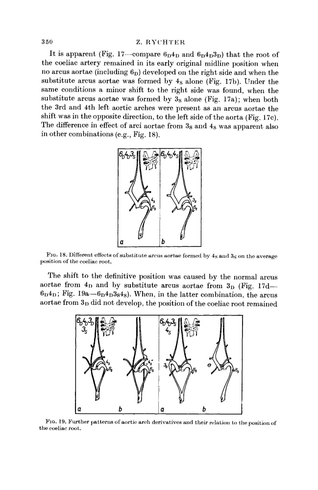
The difference in effect of arci aortae from 3 S and 4 S was apparent also
in other combinations (e.g., Fig. 18).
FIG. 18. Different effects of substitute arcus aortae formed by 4s and 3s on the average
position of the coeliac root.
The shift to the definitive position was caused by the normal arcus
aortae from 4 D and by substitute arcus aortae from 3 D (Fig. 17d—
6 D 4 D ; Fig. 19a—6 D 4 D 3 S 4 S ). When, in the latter combination, the arcus
aortae from 3 D did not develop, the position of the coeliac root remained
FIG. 19. Further patterns of aortic arch derivatives and their relation to the position of
the coeliac root.
Z. RYCHTER
It is apparent (Fig. 17—compare 6 D 4 D and 6 D 4 D 3 D ) that the root of
the coeliac artery remained in its early original midline position when
no arcus aortae (including 6 D ) developed on the right side and when the
substitute arcus aortae was formed by 4 S alone (Fig. 17b). Under the
same conditions a minor shift to the right side was found, when the
substitute arcus aortae was formed by 3 S alone (Fig. 17a); when both
the 3rd and 4th left aortic arches were present as an arcus aortae the
shift was in the opposite direction, to the left side of the aorta (Fig. 17c).
The difference in effect of arci aortae from 3 S and 4 S was apparent also
in other combinations (e.g., Fig. 18).
FIG. 18. Different effects of substitute arcus aortae formed by 4s and 3s on the average
position of the coeliac root.
The shift to the definitive position was caused by the normal arcus
aortae from 4 D and by substitute arcus aortae from 3 D (Fig. 17d—
6 D 4 D ; Fig. 19a—6 D 4 D 3 S 4 S ). When, in the latter combination, the arcus
aortae from 3 D did not develop, the position of the coeliac root remained
FIG. 19. Further patterns of aortic arch derivatives and their relation to the position of
the coeliac root.