

E X P E R I M E N T S ON E M B R Y O N I C A O R T I C A R C H E S 343
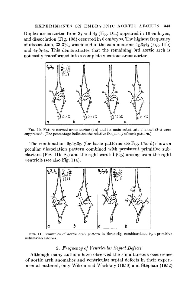
Duplex arcus aortae from 3s and 4 S (Fig. 10a) appeared in 10 embryos,
and dissociation (Fig. lOd) occurred in 8 embryos. The highest frequency
of dissociation, 33-3%, was found in the combinations 4 D 3s4 s (Fig. l i b )
and 4 D 3D4S· This demonstrates that the remaining 3rd aortic arch is
not easily transformed into a complete vicarious arcus aortae.
FIG. 10. Future normal arcus aortae (4D) and its main substitute channel (3D) were
suppressed. (The percentage indicates the relative frequency of each pattern.)
The combination 6 D 4 D 3 D (for basic patterns see Fig. 17a-d) shows a
peculiar dissociation pattern combined with persistent primitive subclavians (Fig. llb-S p ) and the right carotid (CD) arising from the right
ventricle (see also Fig. 11a).
FIG. 11. Examples of aortic arch pattern in three-clip combinations. tt p ^primitive
subclavian arteries.
2. Frequency of Ventricular Septal Defects
Although many authors have observed the simultaneous occurrence
of aortic arch anomalies and ventricular septal defects in their experimental material, only Wilson and Warkany (1950) and Stephan (1952)
Duplex arcus aortae from 3s and 4 S (Fig. 10a) appeared in 10 embryos,
and dissociation (Fig. lOd) occurred in 8 embryos. The highest frequency
of dissociation, 33-3%, was found in the combinations 4 D 3s4 s (Fig. l i b )
and 4 D 3D4S· This demonstrates that the remaining 3rd aortic arch is
not easily transformed into a complete vicarious arcus aortae.
FIG. 10. Future normal arcus aortae (4D) and its main substitute channel (3D) were
suppressed. (The percentage indicates the relative frequency of each pattern.)
The combination 6 D 4 D 3 D (for basic patterns see Fig. 17a-d) shows a
peculiar dissociation pattern combined with persistent primitive subclavians (Fig. llb-S p ) and the right carotid (CD) arising from the right
ventricle (see also Fig. 11a).
FIG. 11. Examples of aortic arch pattern in three-clip combinations. tt p ^primitive
subclavian arteries.
2. Frequency of Ventricular Septal Defects
Although many authors have observed the simultaneous occurrence
of aortic arch anomalies and ventricular septal defects in their experimental material, only Wilson and Warkany (1950) and Stephan (1952)