

342
Z. RYCHTER
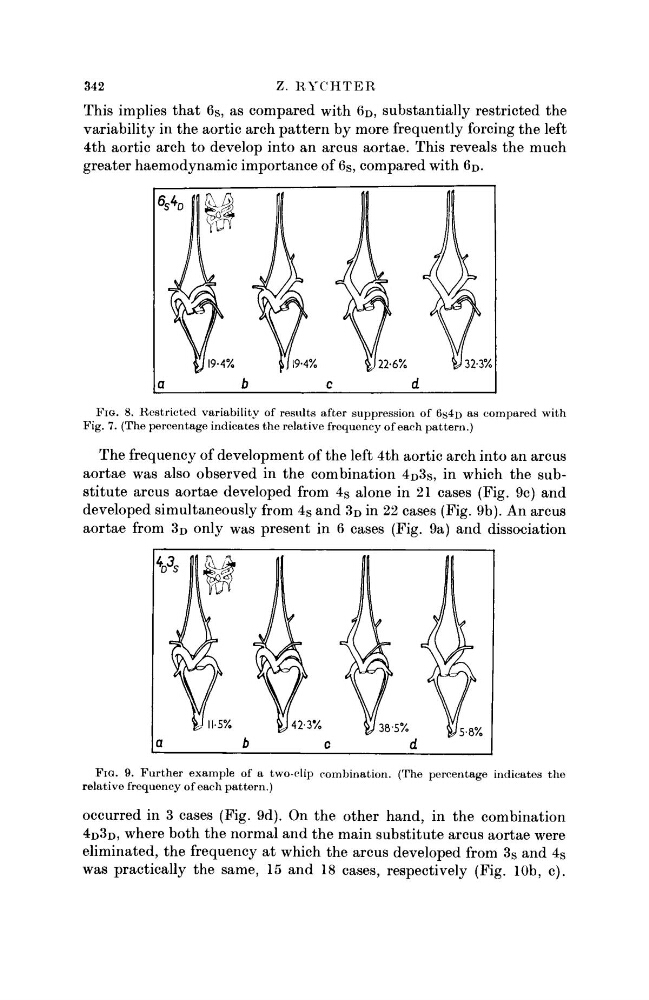
This implies that 6s, as compared with 6D, substantially restricted the
variability in the aortic arch pattern by more frequently forcing the left
4th aortic arch to develop into an arcus aortae. This reveals the much
greater haemodynamic importance of 6 S , compared with 6 D .
FIG. 8. Restricted variability of results after suppression of 6$4D as compared with
Fig. 7. (The percentage indicates the relative frequency of each pattern.)
The frequency of development of the left 4th aortic arch into an arcus
aortae was also observed in the combination 4 D 3s, in which the substitute arcus aortae developed from 4 S alone in 21 cases (Fig. 9c) and
developed simultaneously from 4 S and 3 D in 22 cases (Fig. 9b). An arcus
aortae from 3D only was present in 6 cases (Fig. 9a) and dissociation
FIG. 9. Further example of a two-clip combination. (The percentage indicates the
relative frequency of each pattern.)
occurred in 3 cases (Fig. 9d). On the other hand, in the combination
4D3D, where both the normal and the main substitute arcus aortae were
eliminated, the frequency at which the arcus developed from 3 S and 4 S
was practically the same, 15 and 18 cases, respectively (Fig. 10b, c).
Z. RYCHTER
This implies that 6s, as compared with 6D, substantially restricted the
variability in the aortic arch pattern by more frequently forcing the left
4th aortic arch to develop into an arcus aortae. This reveals the much
greater haemodynamic importance of 6 S , compared with 6 D .
FIG. 8. Restricted variability of results after suppression of 6$4D as compared with
Fig. 7. (The percentage indicates the relative frequency of each pattern.)
The frequency of development of the left 4th aortic arch into an arcus
aortae was also observed in the combination 4 D 3s, in which the substitute arcus aortae developed from 4 S alone in 21 cases (Fig. 9c) and
developed simultaneously from 4 S and 3 D in 22 cases (Fig. 9b). An arcus
aortae from 3D only was present in 6 cases (Fig. 9a) and dissociation
FIG. 9. Further example of a two-clip combination. (The percentage indicates the
relative frequency of each pattern.)
occurred in 3 cases (Fig. 9d). On the other hand, in the combination
4D3D, where both the normal and the main substitute arcus aortae were
eliminated, the frequency at which the arcus developed from 3 S and 4 S
was practically the same, 15 and 18 cases, respectively (Fig. 10b, c).