

E X P E R I M E N T S ON E M B R Y O N I C A O T I C A R C H E S
341
aortic arch. In some cases it was even transposed to the right ventricle,
the narrow pulmonary arteries being its dorsal branches (Fig. 6b).
This very important phenomenon will be analysed in Section II, B, 3.
1. Substitution of the Suppressed Arcus Aortae (4 D )
Because of the large variability in aortic arch arrangements after
suppression of 4 D together with one or two other aortic arches, a large
amount of material was used in the experiments—about 50 embryos for
each combination (Rychter, 1961b).
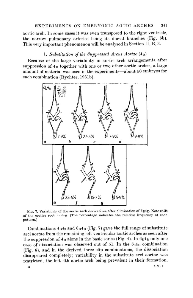
FIG. 7. Variability of the aortic arch derivatives after elimination of 6D4D· Note shift
of the coeliac root in e-g. (The percentage indicates the relative frequency of each
pattern.)
Combinations 4 D 4 S and 6 D 4 D (Fig. 7) gave the full range of substitute
arci aortae from the remaining left ventricular aortic arches as seen after
the suppression of 4 D alone in the basic series (Fig. 4). In 6 D 4 D only one
case of dissociation was observed out of 51. In the 6 S 4 D combination
(Fig. 8), and in the derived three-clip combinations, the dissociation
disappeared completely; variability in the substitute arci aortae was
restricted, the left 4th aortic arch being prevalent in their formation.
M
A . M . 2
341
aortic arch. In some cases it was even transposed to the right ventricle,
the narrow pulmonary arteries being its dorsal branches (Fig. 6b).
This very important phenomenon will be analysed in Section II, B, 3.
1. Substitution of the Suppressed Arcus Aortae (4 D )
Because of the large variability in aortic arch arrangements after
suppression of 4 D together with one or two other aortic arches, a large
amount of material was used in the experiments—about 50 embryos for
each combination (Rychter, 1961b).
FIG. 7. Variability of the aortic arch derivatives after elimination of 6D4D· Note shift
of the coeliac root in e-g. (The percentage indicates the relative frequency of each
pattern.)
Combinations 4 D 4 S and 6 D 4 D (Fig. 7) gave the full range of substitute
arci aortae from the remaining left ventricular aortic arches as seen after
the suppression of 4 D alone in the basic series (Fig. 4). In 6 D 4 D only one
case of dissociation was observed out of 51. In the 6 S 4 D combination
(Fig. 8), and in the derived three-clip combinations, the dissociation
disappeared completely; variability in the substitute arci aortae was
restricted, the left 4th aortic arch being prevalent in their formation.
M
A . M . 2