

666
H O N O R
B .
F E L L
A N D
L .
M . R I N A L D I N I
surrounded by a wreath of vacuolated cells. If the edge of the old
keratin were taken in a pair of fine forceps, it was found to be loose and
unlike that of the controls, could be lifted cleanly away like a lid,
leaving the epithelium intact; no more keratin was formed. With more
prolonged cultivation in vitamin A-containing medium, scattered
secretory cells usually appeared in the epidermis and sometimes areas
of true mucous transformation, but the capacity for metaplasia was
much less than in the 13-day skin.
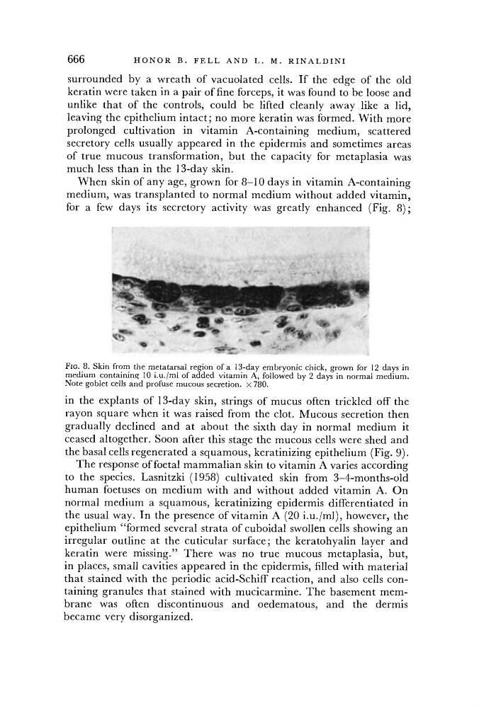
When skin of any age, grown for 8-10 days in vitamin A-containing
medium, was transplanted to normal medium without added vitamin,
for a few days its secretory activity was greatly enhanced (Fig. 8 ) ;
FIG. 8. Skin from the metatarsal region of a 13-day embryonic chick, grown for 12 days in
medium containing 10 i.u./ml of added vitamin A, followed by 2 days in normal medium.
Note goblet cells and profuse mucous secretion. X 780.
in the explants of 13-day skin, strings of mucus often trickled off the
rayon square when it was raised from the clot. Mucous secretion then
gradually declined and at about the sixth day in normal medium it
ceased altogether. Soon after this stage the mucous cells were shed and
the basal cells regenerated a squamous, keratinizing epithelium (Fig. 9).
The response of foetal mammalian skin to vitamin A varies according
to the species. Lasnitzki (1958) cultivated skin from 3-4-months-old
human foetuses on medium with and without added vitamin A. On
normal medium a squamous, keratinizing epidermis differentiated in
the usual way. In the presence of vitamin A (20 i.u./ml), however, the
epithelium "formed several strata of cuboidal swollen cells showing an
irregular outline at the cuticular surface; the keratohyalin layer and
keratin were missing." There was no true mucous metaplasia, but,
in places, small cavities appeared in the epidermis, filled with material
that stained with the periodic acid-Schiff reaction, and also cells containing granules that stained with mucicarmine. The basement membrane was often discontinuous and oedematous, and the dermis
became very disorganized.