

17.
A C T I O N S
O F
V I T A M I N S
A A N D
C
665
to excess of vitamin A in a similar way (Fell, 1957). For this work the
scaley skin from the metatarsus and digits of 13- and 18-day chick
embryos were used. They were grown on squares of rayon cloth on a
plasma and embryo extract clot by Shaffer's modification of the watchglass method (Shaffer, 1956).
The epidermis of the 13-day skin was well differentiated though not
yet keratinized. During cultivation, the scales became more prominent
and keratinization progressed so that the living explants became more
opaque. In the presence of excess vitamin A (10 i.u./ml), however, the
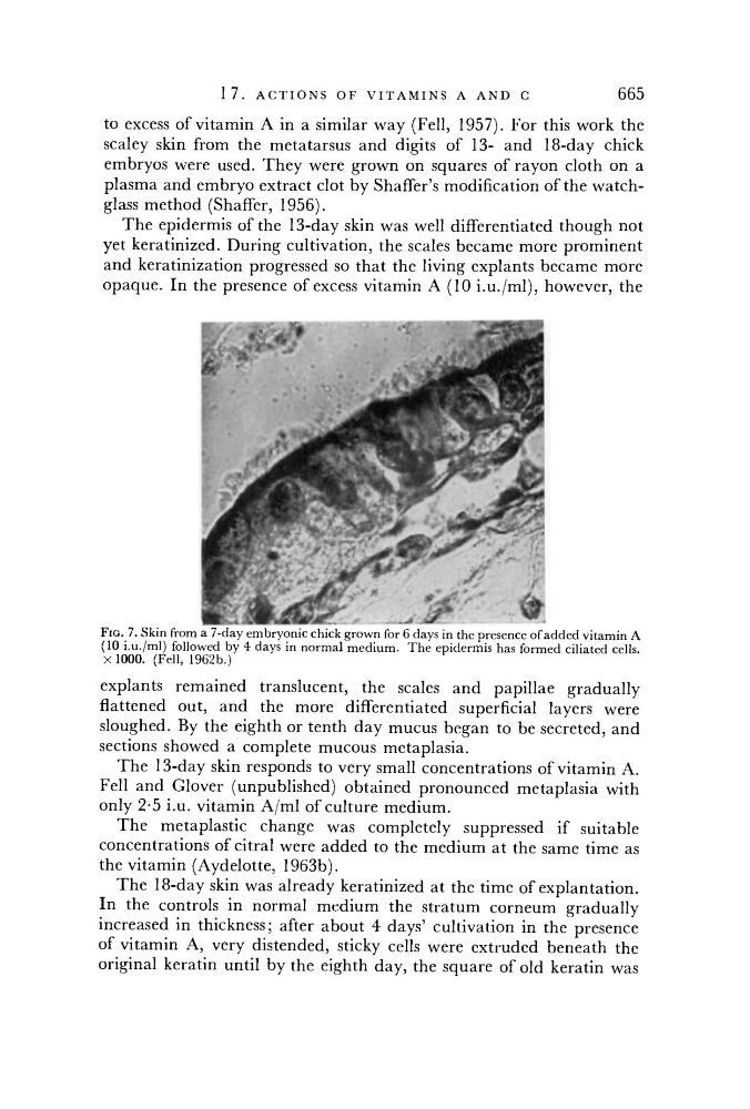
FIG. 7. Skin from a 7-day embryonic chick grown for 6 days in the presence of added vitamin A
(10 i.u./ml) followed by 4 days in normal medium. The epidermis has formed ciliated cells.
X 1000. (Fell, 1962b.)
explants remained translucent, the scales and papillae gradually
flattened out, and the more differentiated superficial layers were
sloughed. By the eighth or tenth day mucus began to be secreted, and
sections showed a complete mucous metaplasia.
The 13-day skin responds to very small concentrations of vitamin A.
Fell and Glover (unpublished) obtained pronounced metaplasia with
only 2-5 i.u. vitamin A/ml of culture medium.
The metaplastic change was completely suppressed if suitable
concentrations of citral were added to the medium at the same time as
the vitamin (Aydelotte, 1963b).
The 18-day skin was already keratinized at the time of explantation.
In the controls in normal medium the stratum corneum gradually
increased in thickness; after about 4 days' cultivation in the presence
of vitamin A, very distended, sticky cells were extruded beneath the
original keratin until by the eighth day, the square of old keratin was