

664
H O N O R
B . F E L L
A N D
L .
M . R I N A L D I N I
directly on clots in watch-glass cultures, but many were cultivated
in large hanging-drop preparations; in these the epidermis rolled
up into a cyst which keratinized in the controls and secreted mucin
in the presence of the vitamin. One day, during a routine examination
of these hanging-drop cultures under the microscope, a loose cell in
one of the cysts of the vitamin A-treated series was seen to be spinning
round in a peculiar way. When the culture was examined under a
higher magnification, an actively beating, ciliated cell was seen in the
wall of the cyst, and it was this ciliary beat that was responsible for the
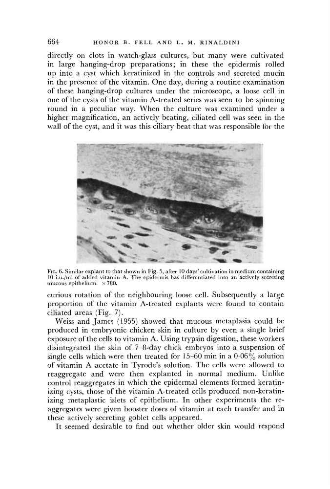
FIG. 6. Similar explant to that shown in Fig. 5, after 10 days' cultivation in medium containing
10 i.u./ml of added vitamin A. The epidermis has differentiated into an actively secreting
mucous epithelium, x 780.
curious rotation of the neighbouring loose cell. Subsequently a large
proportion of the vitamin A-treated explants were found to contain
ciliated areas (Fig. 7).
Weiss and James (1955) showed that mucous metaplasia could be
produced in embryonic chicken skin in culture by even a single brief
exposure ofthe cells to vitamin A. Using trypsin digestion, these workers
disintegrated the skin of 7-8-day chick embryos into a suspension of
single cells which were then treated for 15-60 min in a 0-06% solution
of vitamin A acetate in Tyrode's solution. The cells were allowed to
reaggregate and were then explanted in normal medium. Unlike
control reaggregates in which the epidermal elements formed keratinizing cysts, those of the vitamin A-treated cells produced non-keratinizing metaplastic islets of epithelium. In other experiments the reaggregates were given booster doses of vitamin at each transfer and in
these actively secreting goblet cells appeared.
It seemed desirable to find out whether older skin would respond