

17.
A C T I O N S
O F V I T A M I N S
A
A N D
C
667
When such explants were transplanted to normal medium, the
mucous changes were not accentuated as they are in embryonic
chicken skin transplanted from vitamin A-containing to normal
medium. Instead, the entire epithelium including the basal layer, was
sloughed, and new squamous, keratinizing epithelium regenerated
from the hair follicles.
On the other hand, the dorsal skin of foetal mice and rats when
exposed to vitamin A under similar conditions, does not undergo
mucous metaplasia or cease to keratinize (New, 1963). Even when
skin from a very early (13-day) rat embryo is treated with a high
dose of the vitamin (20 i.u./ml), a squamous keratinizing epidermis
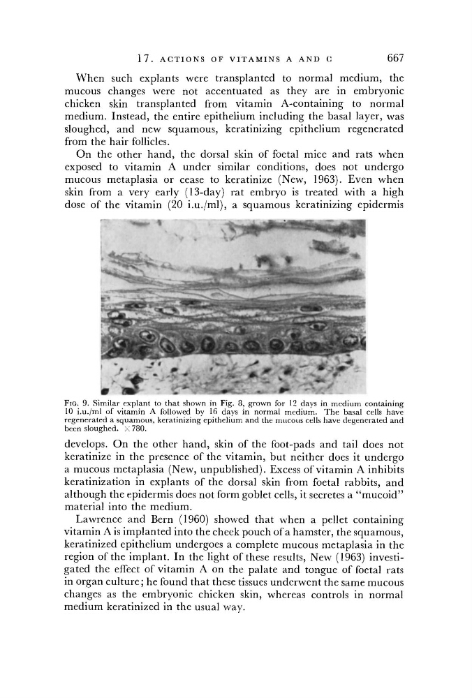
FIG. 9. Similar explant to that shown in Fig. 8, grown for 12 days in medium containing
10 i.u./ml of vitamin A followed by 16 days in normal medium. The basal cells have
regenerated a squamous, keratinizing epithelium and the mucous cells have degenerated and
been sloughed, x 780.
develops. On the other hand, skin of the foot-pads and tail does not
keratinize in the presence of the vitamin, but neither does it undergo
a mucous metaplasia (New, unpublished). Excess of vitamin A inhibits
keratinization in explants of the dorsal skin from foetal rabbits, and
although the epidermis does not form goblet cells, it secretes a "mucoid"
material into the medium.
Lawrence and Bern (1960) showed that when a pellet containing
vitamin A is implanted into the cheek pouch of a hamster, the squamous,
keratinized epithelium undergoes a complete mucous metaplasia in the
region of the implant. In the light of these results, New (1963) investigated the effect of vitamin A on the palate and tongue of foetal rats
in organ culture; he found that these tissues underwent the same mucous
changes as the embryonic chicken skin, whereas controls in normal
medium keratinized in the usual way.