

104
3. Benzenoids
(1966a) separated the pigment from the normal naphthazarins and juglones
by column chromatography on deactivated silica gel. Purification was
achieved by tic and crystallization. Moore et al (1968) determined its
OH
Ο
OH
Ο
47
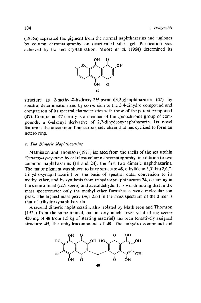
structure as 2-methyl-8-hydroxy-2//-pyrano[3,2-g]naphthazarin (47) by
spectral determination and by conversion to the 3,4-dihydro compound and
comparison of its spectral characteristics with those of the parent compound
(47). Compound 47 clearly is a member of the spinochrome group of compounds, a 6-alkenyl derivative of 2,7-dihydroxynaphthazarin. Its novel
feature is the uncommon four-carbon side chain that has cyclized to form an
hetero ring.
e. The Dimeric
Naphthazarins
Mathieson and Thomson (1971) isolated from the shells of the sea urchin
Spatangus purpureus by cellulose column chromatography, in addition to two
common naphthazarins (11 and 24), the first two dimeric naphthazarins.
The major pigment was shown to have structure 48, ethylidene-3,3'-bis(2,6,7trihydroxynaphthazarin) on the basis of spectral data, conversion to its
methyl ether, and by synthesis from trihydroxynaphthazarin 24, occurring in
the same animal {vide supra) and acetaldehyde. It is worth noting that in the
mass spectrometer only the methyl ether furnishes a weak molecular ion
peak. The highest mass peak {m\e 238) in the mass spectrum of the dimer is
that of trihydroxynaphthazarin.
A second dimeric naphthazarin, also isolated by Mathieson and Thomson
(1971) from the same animal, but in very much lower yield (3 mg versus
420 mg of 48 from 1.5 kg of starting material) has been tentatively assigned
structure 49, the anhydrocompound of 48. The anhydro compound did
OH
Ο
Ο
OH
ΗΟ Ν ^Λ χ ^\^ΟΗ HO N J\ s J^OH
OH
Ο
Ο
OH
48
3. Benzenoids
(1966a) separated the pigment from the normal naphthazarins and juglones
by column chromatography on deactivated silica gel. Purification was
achieved by tic and crystallization. Moore et al (1968) determined its
OH
Ο
OH
Ο
47
structure as 2-methyl-8-hydroxy-2//-pyrano[3,2-g]naphthazarin (47) by
spectral determination and by conversion to the 3,4-dihydro compound and
comparison of its spectral characteristics with those of the parent compound
(47). Compound 47 clearly is a member of the spinochrome group of compounds, a 6-alkenyl derivative of 2,7-dihydroxynaphthazarin. Its novel
feature is the uncommon four-carbon side chain that has cyclized to form an
hetero ring.
e. The Dimeric
Naphthazarins
Mathieson and Thomson (1971) isolated from the shells of the sea urchin
Spatangus purpureus by cellulose column chromatography, in addition to two
common naphthazarins (11 and 24), the first two dimeric naphthazarins.
The major pigment was shown to have structure 48, ethylidene-3,3'-bis(2,6,7trihydroxynaphthazarin) on the basis of spectral data, conversion to its
methyl ether, and by synthesis from trihydroxynaphthazarin 24, occurring in
the same animal {vide supra) and acetaldehyde. It is worth noting that in the
mass spectrometer only the methyl ether furnishes a weak molecular ion
peak. The highest mass peak {m\e 238) in the mass spectrum of the dimer is
that of trihydroxynaphthazarin.
A second dimeric naphthazarin, also isolated by Mathieson and Thomson
(1971) from the same animal, but in very much lower yield (3 mg versus
420 mg of 48 from 1.5 kg of starting material) has been tentatively assigned
structure 49, the anhydrocompound of 48. The anhydro compound did
OH
Ο
Ο
OH
ΗΟ Ν ^Λ χ ^\^ΟΗ HO N J\ s J^OH
OH
Ο
Ο
OH
48