

Chabet Dis, Refes, Varó, Hontoria, Amat and Navarro
2
of Artemia from Sebkha Ezmoul in the Eastern part of Algeria
(Amarouayache and Kara 2015). Ghomari (2013) analysed
the fatty acid profile of six populations (Ezzamoul, Bethioua,
Relizane, El Golea, Adrar and Melghir) belonging to different
inland biotopes (East, West and South of Algeria).
As part of a broader study on the exploration of new
Artemia populations from Algeria, the biometry, hatching
characteristics and fatty acid profile of three Artemia
populations was determined. These three parameters
are used in the quality evaluation of Artemia populations,
according to Lavens and Sorgeloos (1996). These findings
expand and complement existing information.
Materials and methods
Sample collection
Artemia cyst samples were collected directly from
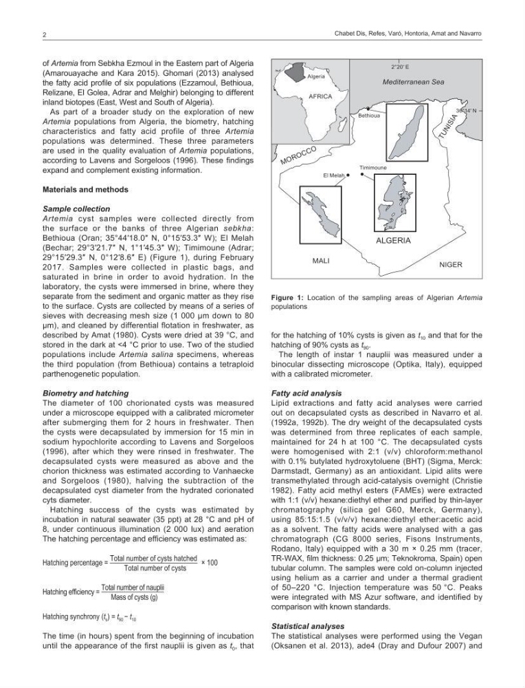
the surface or the banks of three Algerian sebkha:
Bethioua (Oran; 35°44′18.0″ N, 0°15′53.3″ W); El Melah
(Bechar; 29°3′21.7″ N, 1°1′45.3″ W); Timimoune (Adrar;
29°15′29.3″ N, 0°12′8.6″ E) (Figure 1), during February
2017. Samples were collected in plastic bags, and
saturated in brine in order to avoid hydration. In the
laboratory, the cysts were immersed in brine, where they
separate from the sediment and organic matter as they rise
to the surface. Cysts are collected by means of a series of
sieves with decreasing mesh size (1 000 µm down to 80
µm), and cleaned by differential flotation in freshwater, as
described by Amat (1980). Cysts were dried at 39 °C, and
stored in the dark at <4 °C prior to use. Two of the studied
populations include Artemia salina specimens, whereas
the third population (from Bethioua) contains a tetraploid
parthenogenetic population.
Biometry and hatching
The diameter of 100 chorionated cysts was measured
under a microscope equipped with a calibrated micrometer
after submerging them for 2 hours in freshwater. Then
the cysts were decapsulated by immersion for 15 min in
sodium hypochlorite according to Lavens and Sorgeloos
(1996), after which they were rinsed in freshwater. The
decapsulated cysts were measured as above and the
chorion thickness was estimated according to Vanhaecke
and Sorgeloos (1980), halving the subtraction of the
decapsulated cyst diameter from the hydrated corionated
cyts diameter.
Hatching success of the cysts was estimated by
incubation in natural seawater (35 ppt) at 28 °C and pH of
8, under continuous illumination (2 000 lux) and aeration
The hatching percentage and efficiency was estimated as:
Hatching percentage =
Total number of cysts hatched
Total number of cysts
× 100
Hatching efficiency =
Total number of nauplii
Mass of cysts (g)
Hatching synchrony (t s ) = t 90 − t 10
The time (in hours) spent from the beginning of incubation
until the appearance of the first nauplii is given as t 0 , that
for the hatching of 10% cysts is given as t 10 and that for the
hatching of 90% cysts as t 90 .
The length of instar 1 nauplii was measured under a
binocular dissecting microscope (Optika, Italy), equipped
with a calibrated micrometer.
Fatty acid analysis
Lipid extractions and fatty acid analyses were carried
out on decapsulated cysts as described in Navarro et al.
(1992a, 1992b). The dry weight of the decapsulated cysts
was determined from three replicates of each sample,
maintained for 24 h at 100 °C. The decapsulated cysts
were homogenised with 2:1 (v/v) chloroform:methanol
with 0.1% butylated hydroxytoluene (BHT) (Sigma, Merck:
Darmstadt, Germany) as an antioxidant. Lipid alits were
transmethylated through acid-catalysis overnight (Christie
1982). Fatty acid methyl esters (FAMEs) were extracted
with 1:1 (v/v) hexane:diethyl ether and purified by thin-layer
chromatography (silica gel G60, Merck, Germany),
using 85:15:1.5 (v/v/v) hexane:diethyl ether:acetic acid
as a solvent. The fatty acids were analysed with a gas
chromatograph (CG 8000 series, Fisons Instruments,
Rodano, Italy) equipped with a 30 m × 0.25 mm (tracer,
TR-WAX, film thickness: 0.25 µm; Teknokroma, Spain) open
tubular column. The samples were cold on-column injected
using helium as a carrier and under a thermal gradient
of 50–220 °C. Injection temperature was 50 °C. Peaks
were integrated with MS Azur software, and identified by
comparison with known standards.
Statistical analyses
The statistical analyses were performed using the Vegan
(Oksanen et al. 2013), ade4 (Dray and Dufour 2007) and
Mediterranean Sea
M O R O C C O
T U N I S I A
ALGERIA
AFRICA
Algeria
36°14′ N
2°20′ E
NIGER
MALI
Timimoune
El Melah
Bethioua
Figure 1: Location of the sampling areas of Algerian Artemia
populations
2
of Artemia from Sebkha Ezmoul in the Eastern part of Algeria
(Amarouayache and Kara 2015). Ghomari (2013) analysed
the fatty acid profile of six populations (Ezzamoul, Bethioua,
Relizane, El Golea, Adrar and Melghir) belonging to different
inland biotopes (East, West and South of Algeria).
As part of a broader study on the exploration of new
Artemia populations from Algeria, the biometry, hatching
characteristics and fatty acid profile of three Artemia
populations was determined. These three parameters
are used in the quality evaluation of Artemia populations,
according to Lavens and Sorgeloos (1996). These findings
expand and complement existing information.
Materials and methods
Sample collection
Artemia cyst samples were collected directly from
the surface or the banks of three Algerian sebkha:
Bethioua (Oran; 35°44′18.0″ N, 0°15′53.3″ W); El Melah
(Bechar; 29°3′21.7″ N, 1°1′45.3″ W); Timimoune (Adrar;
29°15′29.3″ N, 0°12′8.6″ E) (Figure 1), during February
2017. Samples were collected in plastic bags, and
saturated in brine in order to avoid hydration. In the
laboratory, the cysts were immersed in brine, where they
separate from the sediment and organic matter as they rise
to the surface. Cysts are collected by means of a series of
sieves with decreasing mesh size (1 000 µm down to 80
µm), and cleaned by differential flotation in freshwater, as
described by Amat (1980). Cysts were dried at 39 °C, and
stored in the dark at <4 °C prior to use. Two of the studied
populations include Artemia salina specimens, whereas
the third population (from Bethioua) contains a tetraploid
parthenogenetic population.
Biometry and hatching
The diameter of 100 chorionated cysts was measured
under a microscope equipped with a calibrated micrometer
after submerging them for 2 hours in freshwater. Then
the cysts were decapsulated by immersion for 15 min in
sodium hypochlorite according to Lavens and Sorgeloos
(1996), after which they were rinsed in freshwater. The
decapsulated cysts were measured as above and the
chorion thickness was estimated according to Vanhaecke
and Sorgeloos (1980), halving the subtraction of the
decapsulated cyst diameter from the hydrated corionated
cyts diameter.
Hatching success of the cysts was estimated by
incubation in natural seawater (35 ppt) at 28 °C and pH of
8, under continuous illumination (2 000 lux) and aeration
The hatching percentage and efficiency was estimated as:
Hatching percentage =
Total number of cysts hatched
Total number of cysts
× 100
Hatching efficiency =
Total number of nauplii
Mass of cysts (g)
Hatching synchrony (t s ) = t 90 − t 10
The time (in hours) spent from the beginning of incubation
until the appearance of the first nauplii is given as t 0 , that
for the hatching of 10% cysts is given as t 10 and that for the
hatching of 90% cysts as t 90 .
The length of instar 1 nauplii was measured under a
binocular dissecting microscope (Optika, Italy), equipped
with a calibrated micrometer.
Fatty acid analysis
Lipid extractions and fatty acid analyses were carried
out on decapsulated cysts as described in Navarro et al.
(1992a, 1992b). The dry weight of the decapsulated cysts
was determined from three replicates of each sample,
maintained for 24 h at 100 °C. The decapsulated cysts
were homogenised with 2:1 (v/v) chloroform:methanol
with 0.1% butylated hydroxytoluene (BHT) (Sigma, Merck:
Darmstadt, Germany) as an antioxidant. Lipid alits were
transmethylated through acid-catalysis overnight (Christie
1982). Fatty acid methyl esters (FAMEs) were extracted
with 1:1 (v/v) hexane:diethyl ether and purified by thin-layer
chromatography (silica gel G60, Merck, Germany),
using 85:15:1.5 (v/v/v) hexane:diethyl ether:acetic acid
as a solvent. The fatty acids were analysed with a gas
chromatograph (CG 8000 series, Fisons Instruments,
Rodano, Italy) equipped with a 30 m × 0.25 mm (tracer,
TR-WAX, film thickness: 0.25 µm; Teknokroma, Spain) open
tubular column. The samples were cold on-column injected
using helium as a carrier and under a thermal gradient
of 50–220 °C. Injection temperature was 50 °C. Peaks
were integrated with MS Azur software, and identified by
comparison with known standards.
Statistical analyses
The statistical analyses were performed using the Vegan
(Oksanen et al. 2013), ade4 (Dray and Dufour 2007) and
Mediterranean Sea
M O R O C C O
T U N I S I A
ALGERIA
AFRICA
Algeria
36°14′ N
2°20′ E
NIGER
MALI
Timimoune
El Melah
Bethioua
Figure 1: Location of the sampling areas of Algerian Artemia
populations