

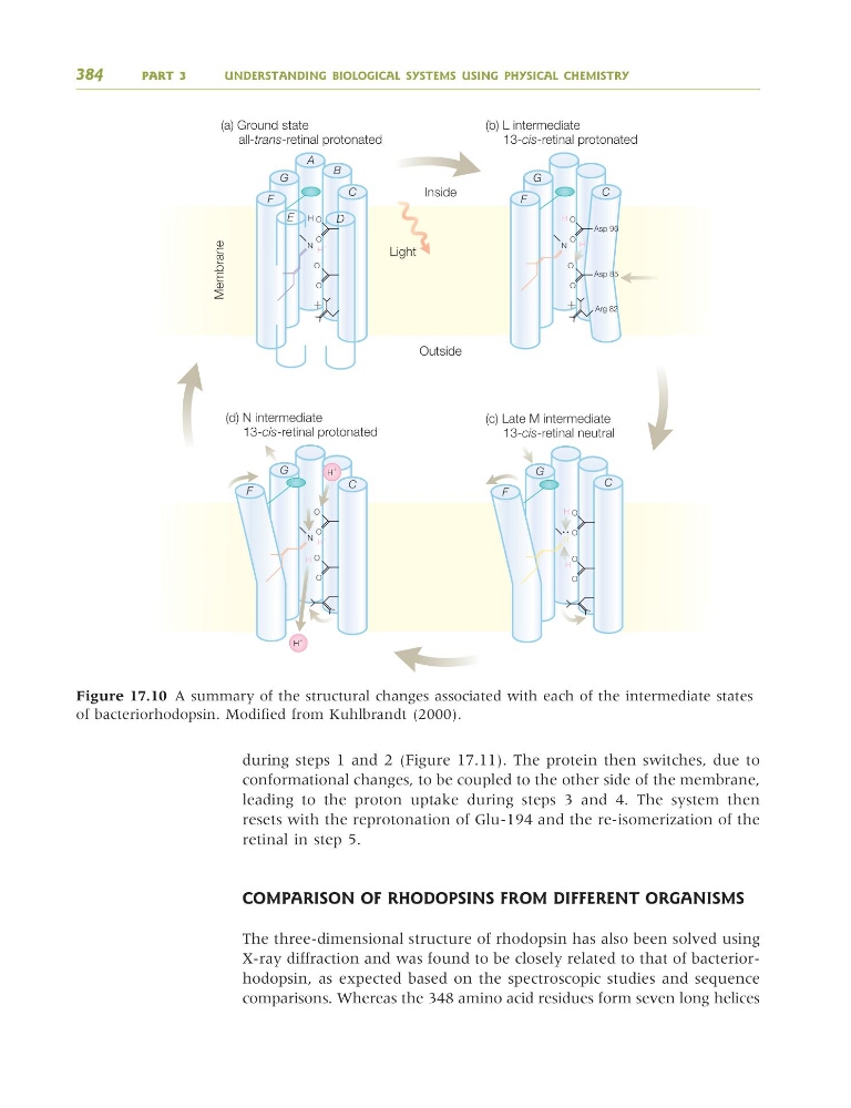
during steps 1 and 2 (Figure 17.11). The protein then switches, due to
conformational changes, to be coupled to the other side of the membrane,
leading to the proton uptake during steps 3 and 4. The system then
resets with the reprotonation of Glu-194 and the re-isomerization of the
retinal in step 5.
COMPARISON OF RHODOPSINS FROM DIFFERENT ORGANISMS
The three-dimensional structure of rhodopsin has also been solved using
X-ray diffraction and was found to be closely related to that of bacteriorhodopsin, as expected based on the spectroscopic studies and sequence
comparisons. Whereas the 348 amino acid residues form seven long helices
384
PART 3
UNDERSTANDING BIOLOGICAL SYSTEMS USING PHYSICAL CHEMISTRY
N
O
(a) Ground state
all-trans-retinal protonated
Inside
Outside
Membrane
Light
ϩ
ϩ
A
B
C
D
E
F
G
C
G
F
C
F
G
(b) L intermediate
13-cis-retinal protonated
H
O
H
ϩ
O
Ϫ
O
N
O
Ϫ
(d) N intermediate
13-cis-retinal protonated
O
H
ϩ
H
ϩ
O
Ϫ
O
N
O
Asp 96
Asp 85
Arg 82
H
O H
ϩ
O
Ϫ
O
H
ϩ
H
ϩ
C
G
F
N
O
(c) Late M intermediate
13-cis-retinal neutral
H
O
H
ϩ
O
O
Figure 17.10 A summary of the structural changes associated with each of the intermediate states
of bacteriorhodopsin. Modified from Kuhlbrandt (2000).
9781405124362_4_017.qxd 4/29/08 10:46 Page 384
conformational changes, to be coupled to the other side of the membrane,
leading to the proton uptake during steps 3 and 4. The system then
resets with the reprotonation of Glu-194 and the re-isomerization of the
retinal in step 5.
COMPARISON OF RHODOPSINS FROM DIFFERENT ORGANISMS
The three-dimensional structure of rhodopsin has also been solved using
X-ray diffraction and was found to be closely related to that of bacteriorhodopsin, as expected based on the spectroscopic studies and sequence
comparisons. Whereas the 348 amino acid residues form seven long helices
384
PART 3
UNDERSTANDING BIOLOGICAL SYSTEMS USING PHYSICAL CHEMISTRY
N
O
(a) Ground state
all-trans-retinal protonated
Inside
Outside
Membrane
Light
ϩ
ϩ
A
B
C
D
E
F
G
C
G
F
C
F
G
(b) L intermediate
13-cis-retinal protonated
H
O
H
ϩ
O
Ϫ
O
N
O
Ϫ
(d) N intermediate
13-cis-retinal protonated
O
H
ϩ
H
ϩ
O
Ϫ
O
N
O
Asp 96
Asp 85
Arg 82
H
O H
ϩ
O
Ϫ
O
H
ϩ
H
ϩ
C
G
F
N
O
(c) Late M intermediate
13-cis-retinal neutral
H
O
H
ϩ
O
O
Figure 17.10 A summary of the structural changes associated with each of the intermediate states
of bacteriorhodopsin. Modified from Kuhlbrandt (2000).
9781405124362_4_017.qxd 4/29/08 10:46 Page 384