

The transition from the dark state to the initial K state happens when
light is absorbed. During the transition from K to L, proton transfer occurs
due to change in the retinal-binding site and a small movement of helix
C that shifts Asp-85 toward the retinal. The proton associated with the
retinal is transferred to Asp-85 as facilitated by Tyr-89 and Asp-212. In
the transition from L to M, the deprotonation causes the retinal to shift,
consequently moving helices F and G. During the M-to-N transition, the
retinal is reprotonated by Asp-96 and a movement of helices F and G
opens a channel that causes Asp-96 to be reprotonated. The protein then
relaxes back to the dark state.
The structure then acts as a proton switch, with one side of the
membrane well coupled to the retinal, leading to the early proton release
CHAPTER 17
SIGNAL TRANSDUCTION
383
402
401
ϩ
H
N
Protonated Schiff base,
all-trans-retinal
(a)
(b)
Thr-89
Tyr-185
Tyr-57
Asp-212
Asp-85
O
O
O
HO
Glu-194
Glu-204
HO
O
OH
C
C
Ϫ
Ϫ
407
406
403
404
405
Arg-82 NH2
NH
HO
O
NH2
C
C
O
OH
H
N
C
O
O
C
ϩ
Ϫ
401
N ¨
Unprotonated Schiff base,
13-cis-retinal
Tyr-185
Trp-86
Tyr-57
Asp-212
Asp-85
O
O
HO
HO
C
Ϫ
407
405
404
Arg-82
Glu-194
Tyr-83
NH2
NH
NH2
C
O
OH
O
C
ϩ
Ϫ
Glu-204
O
O
C
Ϫ
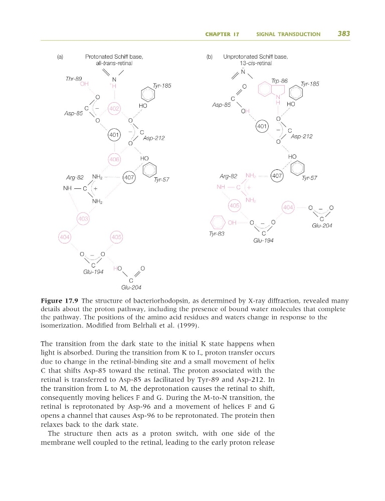
Figure 17.9 The structure of bacteriorhodopsin, as determined by X-ray diffraction, revealed many
details about the proton pathway, including the presence of bound water molecules that complete
the pathway. The positions of the amino acid residues and waters change in response to the
isomerization. Modified from Belrhali et al. (1999).
9781405124362_4_017.qxd 4/29/08 10:46 Page 383
light is absorbed. During the transition from K to L, proton transfer occurs
due to change in the retinal-binding site and a small movement of helix
C that shifts Asp-85 toward the retinal. The proton associated with the
retinal is transferred to Asp-85 as facilitated by Tyr-89 and Asp-212. In
the transition from L to M, the deprotonation causes the retinal to shift,
consequently moving helices F and G. During the M-to-N transition, the
retinal is reprotonated by Asp-96 and a movement of helices F and G
opens a channel that causes Asp-96 to be reprotonated. The protein then
relaxes back to the dark state.
The structure then acts as a proton switch, with one side of the
membrane well coupled to the retinal, leading to the early proton release
CHAPTER 17
SIGNAL TRANSDUCTION
383
402
401
ϩ
H
N
Protonated Schiff base,
all-trans-retinal
(a)
(b)
Thr-89
Tyr-185
Tyr-57
Asp-212
Asp-85
O
O
O
HO
Glu-194
Glu-204
HO
O
OH
C
C
Ϫ
Ϫ
407
406
403
404
405
Arg-82 NH2
NH
HO
O
NH2
C
C
O
OH
H
N
C
O
O
C
ϩ
Ϫ
401
N ¨
Unprotonated Schiff base,
13-cis-retinal
Tyr-185
Trp-86
Tyr-57
Asp-212
Asp-85
O
O
HO
HO
C
Ϫ
407
405
404
Arg-82
Glu-194
Tyr-83
NH2
NH
NH2
C
O
OH
O
C
ϩ
Ϫ
Glu-204
O
O
C
Ϫ
Figure 17.9 The structure of bacteriorhodopsin, as determined by X-ray diffraction, revealed many
details about the proton pathway, including the presence of bound water molecules that complete
the pathway. The positions of the amino acid residues and waters change in response to the
isomerization. Modified from Belrhali et al. (1999).
9781405124362_4_017.qxd 4/29/08 10:46 Page 383