

DETERMINATION OF SECONDARY STRUCTURE USING
CIRCULAR DICHROISM
Chiral molecules can be distinguished based upon their interaction with polarized light. Light can be linearly polarized, in
which case the electromagnetic fields oscillate back and forth
along a line. Alternatively, light can be polarized circularly
when the field rotates as the light propagates, so that when
the light is viewed down the path of travel, the electromagnetic fields are observed to rotate in a circle in either a
right-handed or left-handed direction. For chiral molecules,
right- and left-handed circularly polarized light travels differently in an effect termed circular birefringence. As a result,
chiral molecules show circular dichroism, namely a difference
in their absorbance for left- and right-handed polarized light. In a
circular dichroism spectrum, the wavelength dependence is
measured for the difference in the absorbance for the rightand left-handed polarized light. For proteins, the effect of this
optical activity is evident in a circular dichroism spectrum
that is largely determined by the secondary structure of the
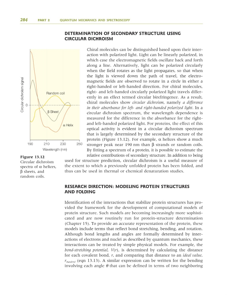
protein (Figure 13.12). For example, α helices show a much
stronger peak near 190 nm than β strands or random coils.
By fitting a spectrum of a protein, it is possible to estimate the
relative contributions of secondary structure. In addition to being
used for structure prediction, circular dichroism is a useful measure of
the extent to which a previously unfolded protein has been folded, and
thus can be used in thermal or chemical denaturation studies.
RESEARCH DIRECTION: MODELING PROTEIN STRUCTURES
AND FOLDING
Identification of the interactions that stabilize protein structures has provided the framework for the development of computational models of
protein structure. Such models are becoming increasingly more sophisticated and are now routinely run for protein-structure determination
(Chapter 15). To provide an accurate representation of the protein, these
models include terms that reflect bond stretching, bending, and rotation.
Although bond lengths and angles are formally determined by interactions of electrons and nuclei as described by quantum mechanics, these
interactions can be treated by simple physical models. For example, the
bond-stretching potential, V(r), is determined by calculating the distance
for each covalent bond, r, and comparing that distance to an ideal 9alue,
r standard (eqn 13.13). A similar expression can be written for the bending
involving each angle θ that can be defined in terms of two neighboring
284
PART 2
QUANTUM MECHANICS AND SPECTROSCOPY
0
190
210
230
250
Random coil
α Helix
β Sheet
Circular dichroism signal
Wavelength (nm)
Figure 13.12
Circular dichroism
spectra of α helices,
β sheets, and
random coils.
9781405124362_4_013.qxd 4/29/08 9:12 Page 284
CIRCULAR DICHROISM
Chiral molecules can be distinguished based upon their interaction with polarized light. Light can be linearly polarized, in
which case the electromagnetic fields oscillate back and forth
along a line. Alternatively, light can be polarized circularly
when the field rotates as the light propagates, so that when
the light is viewed down the path of travel, the electromagnetic fields are observed to rotate in a circle in either a
right-handed or left-handed direction. For chiral molecules,
right- and left-handed circularly polarized light travels differently in an effect termed circular birefringence. As a result,
chiral molecules show circular dichroism, namely a difference
in their absorbance for left- and right-handed polarized light. In a
circular dichroism spectrum, the wavelength dependence is
measured for the difference in the absorbance for the rightand left-handed polarized light. For proteins, the effect of this
optical activity is evident in a circular dichroism spectrum
that is largely determined by the secondary structure of the
protein (Figure 13.12). For example, α helices show a much
stronger peak near 190 nm than β strands or random coils.
By fitting a spectrum of a protein, it is possible to estimate the
relative contributions of secondary structure. In addition to being
used for structure prediction, circular dichroism is a useful measure of
the extent to which a previously unfolded protein has been folded, and
thus can be used in thermal or chemical denaturation studies.
RESEARCH DIRECTION: MODELING PROTEIN STRUCTURES
AND FOLDING
Identification of the interactions that stabilize protein structures has provided the framework for the development of computational models of
protein structure. Such models are becoming increasingly more sophisticated and are now routinely run for protein-structure determination
(Chapter 15). To provide an accurate representation of the protein, these
models include terms that reflect bond stretching, bending, and rotation.
Although bond lengths and angles are formally determined by interactions of electrons and nuclei as described by quantum mechanics, these
interactions can be treated by simple physical models. For example, the
bond-stretching potential, V(r), is determined by calculating the distance
for each covalent bond, r, and comparing that distance to an ideal 9alue,
r standard (eqn 13.13). A similar expression can be written for the bending
involving each angle θ that can be defined in terms of two neighboring
284
PART 2
QUANTUM MECHANICS AND SPECTROSCOPY
0
190
210
230
250
Random coil
α Helix
β Sheet
Circular dichroism signal
Wavelength (nm)
Figure 13.12
Circular dichroism
spectra of α helices,
β sheets, and
random coils.
9781405124362_4_013.qxd 4/29/08 9:12 Page 284