

glutamic acid has been found to have pK A
values shifted by several pH units to maintain its neutral form through interactions
with its environment. The values will also
shift if several ionizable amino acid residues
are near each other. For example, inorganic
nitrogen assimilation in oxygenic photosynthetic organisms has a key step involving the
six-electron reduction of nitrite to ammonia,
which is catalyzed by nitrite reductase. Nitrate
binds to a special cofactor, a siroheme that
is coupled to a four-iron–four-sulfur metal
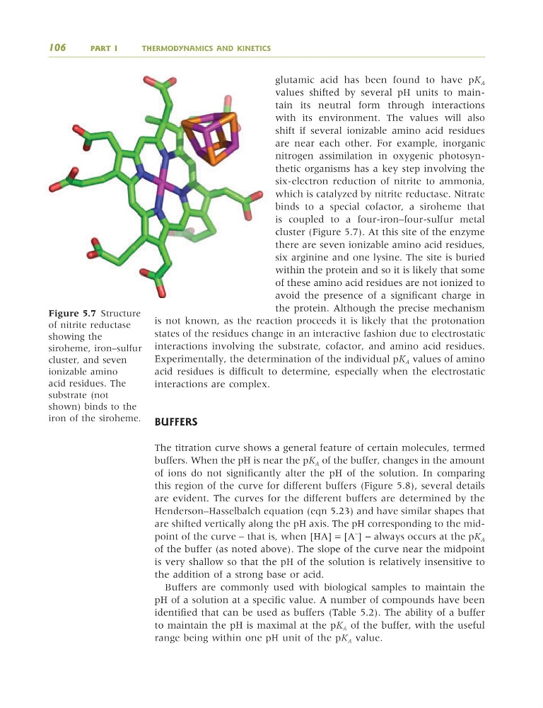
cluster (Figure 5.7). At this site of the enzyme
there are seven ionizable amino acid residues,
six arginine and one lysine. The site is buried
within the protein and so it is likely that some
of these amino acid residues are not ionized to
avoid the presence of a significant charge in
the protein. Although the precise mechanism
is not known, as the reaction proceeds it is likely that the protonation
states of the residues change in an interactive fashion due to electrostatic
interactions involving the substrate, cofactor, and amino acid residues.
Experimentally, the determination of the individual pK A values of amino
acid residues is difficult to determine, especially when the electrostatic
interactions are complex.
BUFFERS
The titration curve shows a general feature of certain molecules, termed
buffers. When the pH is near the pK A of the buffer, changes in the amount
of ions do not significantly alter the pH of the solution. In comparing
this region of the curve for different buffers (Figure 5.8), several details
are evident. The curves for the different buffers are determined by the
Henderson–Hasselbalch equation (eqn 5.23) and have similar shapes that
are shifted vertically along the pH axis. The pH corresponding to the midpoint of the curve – that is, when [HA] = [A
−
] − always occurs at the pK A
of the buffer (as noted above). The slope of the curve near the midpoint
is very shallow so that the pH of the solution is relatively insensitive to
the addition of a strong base or acid.
Buffers are commonly used with biological samples to maintain the
pH of a solution at a specific value. A number of compounds have been
identified that can be used as buffers (Table 5.2). The ability of a buffer
to maintain the pH is maximal at the pK A of the buffer, with the useful
range being within one pH unit of the pK A value.
106
PART I
THERMODYNAMICS AND KINETICS
Figure 5.7 Structure
of nitrite reductase
showing the
siroheme, iron–sulfur
cluster, and seven
ionizable amino
acid residues. The
substrate (not
shown) binds to the
iron of the siroheme.
9781405124362_4_005.qxd 4/29/08 9:08 Page 106
values shifted by several pH units to maintain its neutral form through interactions
with its environment. The values will also
shift if several ionizable amino acid residues
are near each other. For example, inorganic
nitrogen assimilation in oxygenic photosynthetic organisms has a key step involving the
six-electron reduction of nitrite to ammonia,
which is catalyzed by nitrite reductase. Nitrate
binds to a special cofactor, a siroheme that
is coupled to a four-iron–four-sulfur metal
cluster (Figure 5.7). At this site of the enzyme
there are seven ionizable amino acid residues,
six arginine and one lysine. The site is buried
within the protein and so it is likely that some
of these amino acid residues are not ionized to
avoid the presence of a significant charge in
the protein. Although the precise mechanism
is not known, as the reaction proceeds it is likely that the protonation
states of the residues change in an interactive fashion due to electrostatic
interactions involving the substrate, cofactor, and amino acid residues.
Experimentally, the determination of the individual pK A values of amino
acid residues is difficult to determine, especially when the electrostatic
interactions are complex.
BUFFERS
The titration curve shows a general feature of certain molecules, termed
buffers. When the pH is near the pK A of the buffer, changes in the amount
of ions do not significantly alter the pH of the solution. In comparing
this region of the curve for different buffers (Figure 5.8), several details
are evident. The curves for the different buffers are determined by the
Henderson–Hasselbalch equation (eqn 5.23) and have similar shapes that
are shifted vertically along the pH axis. The pH corresponding to the midpoint of the curve – that is, when [HA] = [A
−
] − always occurs at the pK A
of the buffer (as noted above). The slope of the curve near the midpoint
is very shallow so that the pH of the solution is relatively insensitive to
the addition of a strong base or acid.
Buffers are commonly used with biological samples to maintain the
pH of a solution at a specific value. A number of compounds have been
identified that can be used as buffers (Table 5.2). The ability of a buffer
to maintain the pH is maximal at the pK A of the buffer, with the useful
range being within one pH unit of the pK A value.
106
PART I
THERMODYNAMICS AND KINETICS
Figure 5.7 Structure
of nitrite reductase
showing the
siroheme, iron–sulfur
cluster, and seven
ionizable amino
acid residues. The
substrate (not
shown) binds to the
iron of the siroheme.
9781405124362_4_005.qxd 4/29/08 9:08 Page 106