

67
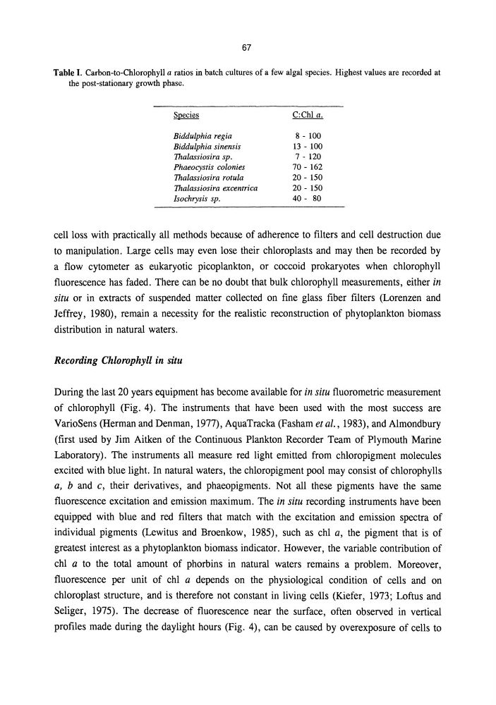
Table I. Carbon-to-Chlorophyll a ratios in batch cultures of a few algal species. Highest values are recorded at
the post-stationary growth phase.
Biddulphia regia
Biddulphia sinensis
Thalassiosira sp.
Phaeocystis colonies
Thalassiosira rotula
Thalassiosira excentrica
Isochrysis sp.
8 - 100
l3 - 100
7 - 120
70 - 162
20 - 150
20 - 150
40 - 80
cell loss with practically all methods because of adherence to filters and cell destruction due
to manipulation. Large cells may even lose their chloroplasts and may then be recorded by
a flow cytometer as eukaryotic picoplankton, or coccoid prokaryotes when chlorophyll
fluorescence has faded. There can be no doubt that bulk chlorophyll measurements, either in
situ or in extracts of suspended matter collected on fine glass fiber filters (Lorenzen and
Jeffrey, 1980), remain a necessity for the realistic reconstruction of phytoplankton biomass
distribution in natural waters.
Recording Chlorophyll in situ
During the last 20 years equipment has become available for in situ fluorometric measurement
of chlorophyll (Fig. 4). The instruments that have been used with the most success are
VarioSens (Herman and Denman, 1977), AquaTracka (Fasham et al., 1983), and Almondbury
(first used by Jim Aitken of the Continuous Plankton Recorder Team of Plymouth Marine
Laboratory). The instruments all measure red light emitted from chloropigment molecules
excited with blue light. In natural waters, the chloropigment pool may consist of chlorophylls
a, band c, their derivatives, and phaeopigments. Not all these pigments have the same
fluorescence excitation and emission maximum. The in situ recording instruments have been
equipped with blue and red filters that match with the excitation and emission spectra of
individual pigments (Lewitus and Broenkow, 1985), such as chI a, the pigment that is of
greatest interest as a phytoplankton biomass indicator. However, the variable contribution of
chI a to the total amount of phorbins in natural waters remains a problem. Moreover,
fluorescence per unit of chI a depends on the physiological condition of cells and on
chloroplast structure, and is therefore not constant in living cells (Kiefer, 1973; Loftus and
Seliger, 1975). The decrease of fluorescence near the surface, often observed in vertical
profiles made during the daylight hours (Fig. 4), can be caused by overexposure of cells to
Table I. Carbon-to-Chlorophyll a ratios in batch cultures of a few algal species. Highest values are recorded at
the post-stationary growth phase.
Biddulphia regia
Biddulphia sinensis
Thalassiosira sp.
Phaeocystis colonies
Thalassiosira rotula
Thalassiosira excentrica
Isochrysis sp.
8 - 100
l3 - 100
7 - 120
70 - 162
20 - 150
20 - 150
40 - 80
cell loss with practically all methods because of adherence to filters and cell destruction due
to manipulation. Large cells may even lose their chloroplasts and may then be recorded by
a flow cytometer as eukaryotic picoplankton, or coccoid prokaryotes when chlorophyll
fluorescence has faded. There can be no doubt that bulk chlorophyll measurements, either in
situ or in extracts of suspended matter collected on fine glass fiber filters (Lorenzen and
Jeffrey, 1980), remain a necessity for the realistic reconstruction of phytoplankton biomass
distribution in natural waters.
Recording Chlorophyll in situ
During the last 20 years equipment has become available for in situ fluorometric measurement
of chlorophyll (Fig. 4). The instruments that have been used with the most success are
VarioSens (Herman and Denman, 1977), AquaTracka (Fasham et al., 1983), and Almondbury
(first used by Jim Aitken of the Continuous Plankton Recorder Team of Plymouth Marine
Laboratory). The instruments all measure red light emitted from chloropigment molecules
excited with blue light. In natural waters, the chloropigment pool may consist of chlorophylls
a, band c, their derivatives, and phaeopigments. Not all these pigments have the same
fluorescence excitation and emission maximum. The in situ recording instruments have been
equipped with blue and red filters that match with the excitation and emission spectra of
individual pigments (Lewitus and Broenkow, 1985), such as chI a, the pigment that is of
greatest interest as a phytoplankton biomass indicator. However, the variable contribution of
chI a to the total amount of phorbins in natural waters remains a problem. Moreover,
fluorescence per unit of chI a depends on the physiological condition of cells and on
chloroplast structure, and is therefore not constant in living cells (Kiefer, 1973; Loftus and
Seliger, 1975). The decrease of fluorescence near the surface, often observed in vertical
profiles made during the daylight hours (Fig. 4), can be caused by overexposure of cells to