

Bacteria
352
(
photoacclimation
PHYSIOLOOY
Water column
t Cell Sorting
mixing
In situ Growth
OPTICS
(DNA, Light Scatter)
Fluorescence ~ Absorption
~
--------~~
T
.. Light Scatter ~ Size,
Bottle Incubations
+
Distributions
t
Eukaryotic phytoplankton
t '-Large sample volume
Picophytoplankton
T
Refractive Index
Beam Attenuation
Immunofluorescence
Oligonucleotide probes
ECOLO~
(Organis;j ,
Grazing ~
Phytoplankton DNA
(cell cycle)
Phytoplankton Fluorescence, Light Scatter
"'~I--__ ~_-=I Biomedical Applications
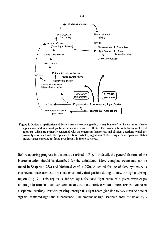
Figure 1. Outline of applications of flow cytometry to oceanography, attempting to reflect the evolution of these
applications and relationships between various research efforts. The major split is between ecological
questions, which are primarily concerned with the organisms themselves, and physical questions, which are
primarily concerned with the optical effects of particles, regardless of their origin or composition. Italics
indicate areas expected to figure prominently in future advances.
Before covering progress in the areas described in Fig. 1 in detail, the general features of the
instrumentation should be described for the uninitiated. More complete treatments can be
found in Shapiro (1988) and Melamed et ai. (1990). A central feature of flow cytometry is
that several measurements are made on an individual particle during its flow through a sensing
region (Fig. 2). This region is defined by a focussed light beam of a given wavelength
(although instruments that can also make electronic particle volume measurements do so in
a separate location). Particles passing through this light beam give rise to two kinds of optical
signals: scattered light and fluorescence. The amount of light scattered from the beam by a
352
(
photoacclimation
PHYSIOLOOY
Water column
t Cell Sorting
mixing
In situ Growth
OPTICS
(DNA, Light Scatter)
Fluorescence ~ Absorption
~
--------~~
T
.. Light Scatter ~ Size,
Bottle Incubations
+
Distributions
t
Eukaryotic phytoplankton
t '-Large sample volume
Picophytoplankton
T
Refractive Index
Beam Attenuation
Immunofluorescence
Oligonucleotide probes
ECOLO~
(Organis;j ,
Grazing ~
Phytoplankton DNA
(cell cycle)
Phytoplankton Fluorescence, Light Scatter
"'~I--__ ~_-=I Biomedical Applications
Figure 1. Outline of applications of flow cytometry to oceanography, attempting to reflect the evolution of these
applications and relationships between various research efforts. The major split is between ecological
questions, which are primarily concerned with the organisms themselves, and physical questions, which are
primarily concerned with the optical effects of particles, regardless of their origin or composition. Italics
indicate areas expected to figure prominently in future advances.
Before covering progress in the areas described in Fig. 1 in detail, the general features of the
instrumentation should be described for the uninitiated. More complete treatments can be
found in Shapiro (1988) and Melamed et ai. (1990). A central feature of flow cytometry is
that several measurements are made on an individual particle during its flow through a sensing
region (Fig. 2). This region is defined by a focussed light beam of a given wavelength
(although instruments that can also make electronic particle volume measurements do so in
a separate location). Particles passing through this light beam give rise to two kinds of optical
signals: scattered light and fluorescence. The amount of light scattered from the beam by a