

19
off very rapidly with increasing scattering angle, and more so for larger particles. At small
scattering angles, i.e. below 5°, N\(a) depends primarily on cell size, whereas at higher
angles, i.e. above 10°, N\(a) is also significantly influenced by the shape and internal structure
of the cell. Small structures, such as intracellular granules, will contribute more to N\(a) at
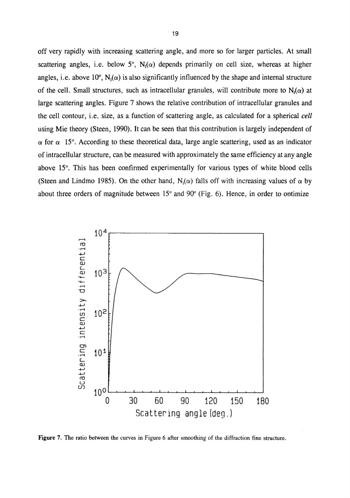
large scattering angles. Figure 7 shows the relative contribution of intracellular granules and
the cell contour, i.e. size, as a function of scattering angle, as calculated for a spherical cell
using Mie theory (Steen, 1990). It can be seen that this contribution is largely independent of
a for a 15°. According to these theoretical data, large angle scattering, used as an indicator
of intracellular structure, can be measured with approximately the same efficiency at any angle
above 15°. This has been confirmed experimentally for various types of white blood cells
(Steen and Lindmo 1985). On the other hand, N\(a) falls off with increasing values of a by
about three orders of magnitude between 15° and 90° (Fig. 6). Hence, in order to ootimize
10 4
r-t
m
-r-!
..,
c::
UJ
L
10 3
UJ
4-r-!
TI
>..,
-r-!
10 2
(J)
c::
OJ
..,
c::
-r-!
OJ
c::
10 1
-r-!
L
UJ
..,
..,
m
u
CfJ
10°
0
30
60
90
120 150 180
Scattering angle (deg.J
Figure 7. The ratio between the curves in Figure 6 after smoothing of the diffraction fine structure.
off very rapidly with increasing scattering angle, and more so for larger particles. At small
scattering angles, i.e. below 5°, N\(a) depends primarily on cell size, whereas at higher
angles, i.e. above 10°, N\(a) is also significantly influenced by the shape and internal structure
of the cell. Small structures, such as intracellular granules, will contribute more to N\(a) at
large scattering angles. Figure 7 shows the relative contribution of intracellular granules and
the cell contour, i.e. size, as a function of scattering angle, as calculated for a spherical cell
using Mie theory (Steen, 1990). It can be seen that this contribution is largely independent of
a for a 15°. According to these theoretical data, large angle scattering, used as an indicator
of intracellular structure, can be measured with approximately the same efficiency at any angle
above 15°. This has been confirmed experimentally for various types of white blood cells
(Steen and Lindmo 1985). On the other hand, N\(a) falls off with increasing values of a by
about three orders of magnitude between 15° and 90° (Fig. 6). Hence, in order to ootimize
10 4
r-t
m
-r-!
..,
c::
UJ
L
10 3
UJ
4-r-!
TI
>..,
-r-!
10 2
(J)
c::
OJ
..,
c::
-r-!
OJ
c::
10 1
-r-!
L
UJ
..,
..,
m
u
CfJ
10°
0
30
60
90
120 150 180
Scattering angle (deg.J
Figure 7. The ratio between the curves in Figure 6 after smoothing of the diffraction fine structure.