

1. Cadet et al. : DNA Modifications Due to Oxidative Damage
285
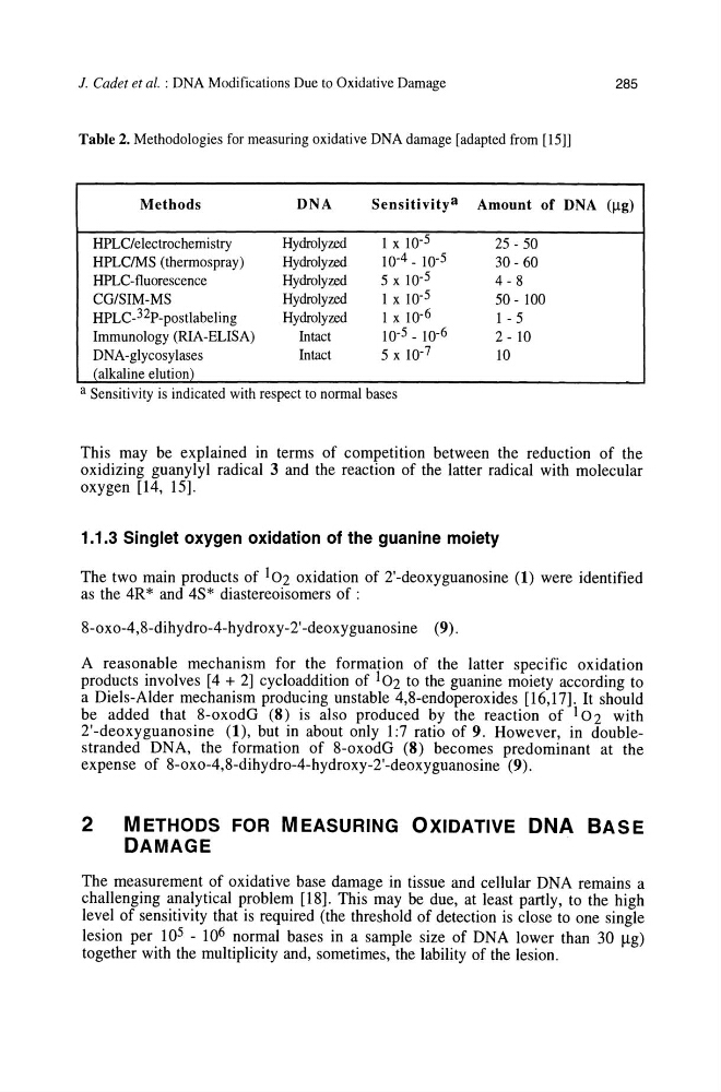
Table 2. Methodologies for measuring oxidative DNA damage [adapted from [15]]
Methods
DNA
Sensitivitya Amount of DNA (Il-g)
HPLC/electrochemistry
Hydrolyzed
I x 10- 5
25 - 50
HPLCIMS (thermospray)
Hydrolyzed
10-4 - 10-5
30 - 60
HPLC-tluorescence
Hydrolyzed
5 x 10- 5
4-8
CG/SIM-MS
Hydrolyzed
I x 10-5
50 - 100
HPLC_32P-postlabeling
Hydrolyzed
I x 10- 6
I - 5
Immunology (RIA-ELISA)
Intact
10-5 _ 10-6
2 - 10
DNA-glycosylases
Intact
5 x 10- 7
10
(alkaline elution)
a Sensitivity is indicated with respect to normal bases
This may be explained in terms of competition between the reduction of the
oxidizing guanylyl radical 3 and the reaction of the latter radical with molecular
oxygen [14, 15].
1.1.3 Singlet oxygen oxidation of the guanine moiety
The two main products of 102 oxidation of 2'-deoxyguanosine (1) were identified
as the 4R* and 4S* diastereoisomers of :
8-oxo-4,8-dihydro-4-hydroxy-2'-deoxyguanosine (9).
A reasonable mechanism for the formation of the latter specific oxidation
products involves [4 + 2] cycloaddition of 102 to the guanine moiety according to
a Diels-Alder mechanism producing unstable 4,8-endoperoxides [16,17]. It should
be added that 8-oxodG (8) is also produced by the reaction of 102 with
2'-deoxyguanosine (1), but in about only 1:7 ratio of 9. However, in doublestranded DNA, the formation of 8-oxodG (8) becomes predominant at the
expense of 8-oxo-4,8-dihydro-4-hydroxy-2'-deoxyguanosine (9).
2
METHODS FOR MEASURING OXIDATIVE DNA BASE
DAMAGE
The measurement of oxidative base damage in tissue and cellular DNA remains a
challenging analytical problem [18]. This may be due, at least partly, to the high
level of sensitivity that is required (the threshold of detection is close to one single
lesion per 10 5 - 10 6 normal bases in a sample size of DNA lower than 30 Ilg)
together with the multiplicity and, sometimes, the lability of the lesion.
285
Table 2. Methodologies for measuring oxidative DNA damage [adapted from [15]]
Methods
DNA
Sensitivitya Amount of DNA (Il-g)
HPLC/electrochemistry
Hydrolyzed
I x 10- 5
25 - 50
HPLCIMS (thermospray)
Hydrolyzed
10-4 - 10-5
30 - 60
HPLC-tluorescence
Hydrolyzed
5 x 10- 5
4-8
CG/SIM-MS
Hydrolyzed
I x 10-5
50 - 100
HPLC_32P-postlabeling
Hydrolyzed
I x 10- 6
I - 5
Immunology (RIA-ELISA)
Intact
10-5 _ 10-6
2 - 10
DNA-glycosylases
Intact
5 x 10- 7
10
(alkaline elution)
a Sensitivity is indicated with respect to normal bases
This may be explained in terms of competition between the reduction of the
oxidizing guanylyl radical 3 and the reaction of the latter radical with molecular
oxygen [14, 15].
1.1.3 Singlet oxygen oxidation of the guanine moiety
The two main products of 102 oxidation of 2'-deoxyguanosine (1) were identified
as the 4R* and 4S* diastereoisomers of :
8-oxo-4,8-dihydro-4-hydroxy-2'-deoxyguanosine (9).
A reasonable mechanism for the formation of the latter specific oxidation
products involves [4 + 2] cycloaddition of 102 to the guanine moiety according to
a Diels-Alder mechanism producing unstable 4,8-endoperoxides [16,17]. It should
be added that 8-oxodG (8) is also produced by the reaction of 102 with
2'-deoxyguanosine (1), but in about only 1:7 ratio of 9. However, in doublestranded DNA, the formation of 8-oxodG (8) becomes predominant at the
expense of 8-oxo-4,8-dihydro-4-hydroxy-2'-deoxyguanosine (9).
2
METHODS FOR MEASURING OXIDATIVE DNA BASE
DAMAGE
The measurement of oxidative base damage in tissue and cellular DNA remains a
challenging analytical problem [18]. This may be due, at least partly, to the high
level of sensitivity that is required (the threshold of detection is close to one single
lesion per 10 5 - 10 6 normal bases in a sample size of DNA lower than 30 Ilg)
together with the multiplicity and, sometimes, the lability of the lesion.