

62
D.R. Ketten
responses were obtained from sources placed on or near the external
meatus. Renaud and Popper (1975) similarly found a response split at
20kHz for sources directed at or behind the jaw in psychophysical localization tests with T. truncatus. Popov and Supin (1990) found minimum
thresholds were associated with stimuli near the external meatus for multiple lower-frequency stimuli. McCormick et al. (1970) did not do specific
frequency versus source location comparisons but commented that they
found a strong cochlear microphonic response for 2 kHz airborne sources
both placed over the meatus and over the jaw.
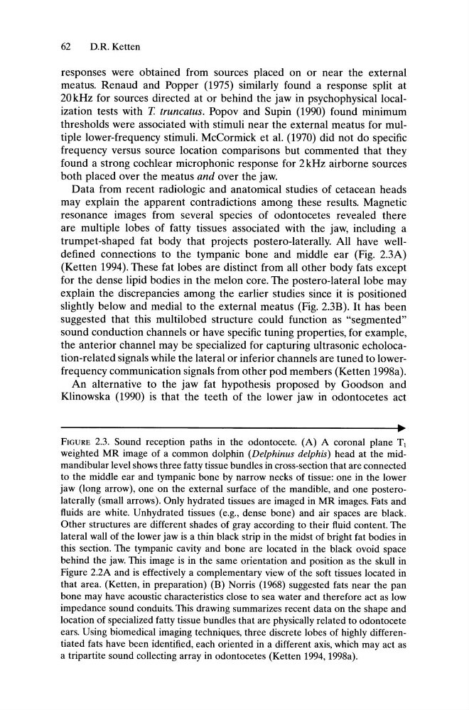
Data from recent radiologic and anatomical studies of cetacean heads
may explain the apparent contradictions among these results. Magnetic
resonance images from several species of odontocetes revealed there
are multiple lobes of fatty tissues associated with the jaw, including a
trumpet-shaped fat body that projects postero-Iaterally. All have welldefined connections to the tympanic bone and middle ear (Fig. 2.3A)
(Ketten 1994). These fat lobes are distinct from all other body fats except
for the dense lipid bodies in the melon core. The postero-Iateral lobe may
explain the discrepancies among the earlier studies since it is positioned
slightly below and medial to the external meatus (Fig. 2.3B). It has been
suggested that this multilobed structure could function as "segmented"
sound conduction channels or have specific tuning properties, for example,
the anterior channel may be specialized for capturing ultrasonic echolocation-related signals while the lateral or inferior channels are tuned to lowerfrequency communication signals from other pod members (Ketten 1998a).
An alternative to the jaw fat hypothesis proposed by Goodson and
Klinowska (1990) is that the teeth of the lower jaw in odontocetes act
FIGURE 2.3. Sound reception paths in the odontocete. (A) A coronal plane T[
weighted MR image of a common dolphin (Delphinus delphis) head at the midmandibular level shows three fatty tissue bundles in cross-section that are connected
to the middle ear and tympanic bone by narrow necks of tissue: one in the lower
jaw (long arrow), one on the external surface of the mandible, and one posterolaterally (small arrows). Only hydrated tissues are imaged in MR images. Fats and
fluids are white. Unhydrated tissues (e.g., dense bone) and air spaces are black.
Other structures are different shades of gray according to their fluid content. The
lateral wall of the lower jaw is a thin black strip in the midst of bright fat bodies in
this section. The tympanic cavity and bone are located in the black ovoid space
behind the jaw. This image is in the same orientation and position as the skull in
Figure 2.2A and is effectively a complementary view of the soft tissues located in
that area. (Ketten, in preparation) (B) Norris (1968) suggested fats near the pan
bone may have acoustic characteristics close to sea water and therefore act as low
impedance sound conduits. This drawing summarizes recent data on the shape and
location of specialized fatty tissue bundles that are physically related to odontocete
ears. Using biomedical imaging techniques, three discrete lobes of highly differentiated fats have been identified, each oriented in a different axis, which may act as
a tripartite sound collecting array in odontocetes (Ketten 1994, 1998a).
D.R. Ketten
responses were obtained from sources placed on or near the external
meatus. Renaud and Popper (1975) similarly found a response split at
20kHz for sources directed at or behind the jaw in psychophysical localization tests with T. truncatus. Popov and Supin (1990) found minimum
thresholds were associated with stimuli near the external meatus for multiple lower-frequency stimuli. McCormick et al. (1970) did not do specific
frequency versus source location comparisons but commented that they
found a strong cochlear microphonic response for 2 kHz airborne sources
both placed over the meatus and over the jaw.
Data from recent radiologic and anatomical studies of cetacean heads
may explain the apparent contradictions among these results. Magnetic
resonance images from several species of odontocetes revealed there
are multiple lobes of fatty tissues associated with the jaw, including a
trumpet-shaped fat body that projects postero-Iaterally. All have welldefined connections to the tympanic bone and middle ear (Fig. 2.3A)
(Ketten 1994). These fat lobes are distinct from all other body fats except
for the dense lipid bodies in the melon core. The postero-Iateral lobe may
explain the discrepancies among the earlier studies since it is positioned
slightly below and medial to the external meatus (Fig. 2.3B). It has been
suggested that this multilobed structure could function as "segmented"
sound conduction channels or have specific tuning properties, for example,
the anterior channel may be specialized for capturing ultrasonic echolocation-related signals while the lateral or inferior channels are tuned to lowerfrequency communication signals from other pod members (Ketten 1998a).
An alternative to the jaw fat hypothesis proposed by Goodson and
Klinowska (1990) is that the teeth of the lower jaw in odontocetes act
FIGURE 2.3. Sound reception paths in the odontocete. (A) A coronal plane T[
weighted MR image of a common dolphin (Delphinus delphis) head at the midmandibular level shows three fatty tissue bundles in cross-section that are connected
to the middle ear and tympanic bone by narrow necks of tissue: one in the lower
jaw (long arrow), one on the external surface of the mandible, and one posterolaterally (small arrows). Only hydrated tissues are imaged in MR images. Fats and
fluids are white. Unhydrated tissues (e.g., dense bone) and air spaces are black.
Other structures are different shades of gray according to their fluid content. The
lateral wall of the lower jaw is a thin black strip in the midst of bright fat bodies in
this section. The tympanic cavity and bone are located in the black ovoid space
behind the jaw. This image is in the same orientation and position as the skull in
Figure 2.2A and is effectively a complementary view of the soft tissues located in
that area. (Ketten, in preparation) (B) Norris (1968) suggested fats near the pan
bone may have acoustic characteristics close to sea water and therefore act as low
impedance sound conduits. This drawing summarizes recent data on the shape and
location of specialized fatty tissue bundles that are physically related to odontocete
ears. Using biomedical imaging techniques, three discrete lobes of highly differentiated fats have been identified, each oriented in a different axis, which may act as
a tripartite sound collecting array in odontocetes (Ketten 1994, 1998a).