

60
D.R. Ketten
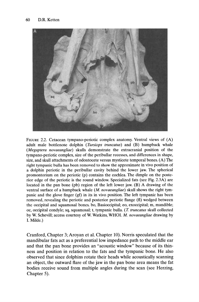
FIGURE 2.2. Cetacean tympano-periotic complex anatomy. Ventral views of (A)
adult male bottlenose dolphin (Tursiops truncatus) and (B) humpback whale
(Megaptera novaeangliae) skulls demonstrate the extracranial position of the
tympano-periotic complex, size of the peribullar recesses, and differences in shape,
size, and skull attachments of odontocete versus mysticete temporal bones. (A) The
right tympanic bulla has been removed to show the approximate in vivo position of
a dolphin periotic in the peribullar cavity behind the lower jaw. The spherical
promontorium on the periotic (p) contains the cochlea. The dimple on the posterior edge of the periotic is the round window. Specialized fats (see Fig. 2.3A) are
located in the pan bone (pb) region of the left lower jaw. (B) A drawing of the
ventral surface of a humpback whale (M. novaeangliae) skull shows the right tympanic and the glove finger (gf) in its in vivo position. The left tympanic has been
removed, revealing the periotic and posterior periotic flange (fl) wedged between
the occipital and squamosal bones. bo, Basioccipital; eo, exoccipital; m, mandible;
oc, occipital condyle; sq, squamosal; t, tympanic bulla. (T truncatus skull collected
by W. Schevill; access courtesy of W. Watkins, WHOI. M. novaeangliae drawing by
I. Milde.)
Cranford, Chapter 3; Aroyan et al. Chapter 10). Norris speculated that the
mandibular fats act as a preferential low impedance path to the middle ear
and that the pan bone provides an "acoustic window" because of its thinness and position in relation to the fats and the tympanic bone. He also
observed that since dolphins rotate their heads while acoustically scanning
an object, the outward flare of the jaw in the pan bone area means the fat
bodies receive sound from multiple angles during the scan (see Herzing,
Chapter 5).
D.R. Ketten
FIGURE 2.2. Cetacean tympano-periotic complex anatomy. Ventral views of (A)
adult male bottlenose dolphin (Tursiops truncatus) and (B) humpback whale
(Megaptera novaeangliae) skulls demonstrate the extracranial position of the
tympano-periotic complex, size of the peribullar recesses, and differences in shape,
size, and skull attachments of odontocete versus mysticete temporal bones. (A) The
right tympanic bulla has been removed to show the approximate in vivo position of
a dolphin periotic in the peribullar cavity behind the lower jaw. The spherical
promontorium on the periotic (p) contains the cochlea. The dimple on the posterior edge of the periotic is the round window. Specialized fats (see Fig. 2.3A) are
located in the pan bone (pb) region of the left lower jaw. (B) A drawing of the
ventral surface of a humpback whale (M. novaeangliae) skull shows the right tympanic and the glove finger (gf) in its in vivo position. The left tympanic has been
removed, revealing the periotic and posterior periotic flange (fl) wedged between
the occipital and squamosal bones. bo, Basioccipital; eo, exoccipital; m, mandible;
oc, occipital condyle; sq, squamosal; t, tympanic bulla. (T truncatus skull collected
by W. Schevill; access courtesy of W. Watkins, WHOI. M. novaeangliae drawing by
I. Milde.)
Cranford, Chapter 3; Aroyan et al. Chapter 10). Norris speculated that the
mandibular fats act as a preferential low impedance path to the middle ear
and that the pan bone provides an "acoustic window" because of its thinness and position in relation to the fats and the tympanic bone. He also
observed that since dolphins rotate their heads while acoustically scanning
an object, the outward flare of the jaw in the pan bone area means the fat
bodies receive sound from multiple angles during the scan (see Herzing,
Chapter 5).