

6. Auditory eNS of Dolphins
275
2. The Ear
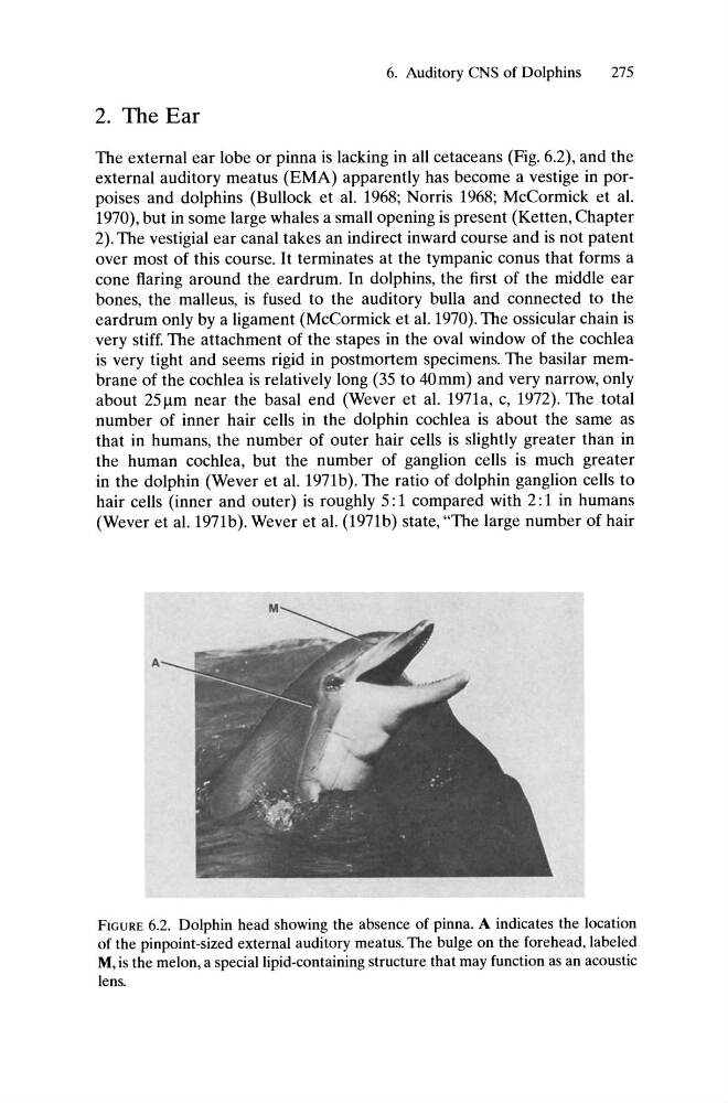
The external ear lobe or pinna is lacking in all cetaceans (Fig. 6.2), and the
external auditory meatus (EMA) apparently has become a vestige in porpoises and dolphins (Bullock et al. 1968; Norris 1968; McCormick et al.
1970), but in some large whales a small opening is present (Ketten, Chapter
2). The vestigial ear canal takes an indirect inward course and is not patent
over most of this course. It terminates at the tympanic conus that forms a
cone flaring around the eardrum. In dolphins, the first of the middle ear
bones, the malleus, is fused to the auditory bulla and connected to the
eardrum only by a ligament (McCormick et al. 1970). The ossicular chain is
very stiff. The attachment of the stapes in the oval window of the cochlea
is very tight and seems rigid in postmortem specimens. The basilar membrane of the cochlea is relatively long (35 to 40mm) and very narrow, only
about 251-Im near the basal end (Wever et al. 1971a, c, 1972). The total
number of inner hair cells in the dolphin cochlea is about the same as
that in humans, the number of outer hair cells is slightly greater than in
the human cochlea, but the number of ganglion cells is much greater
in the dolphin (Wever et al. 1971b). The ratio of dolphin ganglion cells to
hair cells (inner and outer) is roughly 5: 1 compared with 2: 1 in humans
(Wever et al. 1971b). Wever et al. (1971b) state, "The large number of hair
A
FIGURE 6.2. Dolphin head showing the absence of pinna. A indicates the location
of the pinpoint-sized external auditory meatus. The bulge on the forehead, labeled
M, is the melon, a special lipid-containing structure that may function as an acoustic
lens.
275
2. The Ear
The external ear lobe or pinna is lacking in all cetaceans (Fig. 6.2), and the
external auditory meatus (EMA) apparently has become a vestige in porpoises and dolphins (Bullock et al. 1968; Norris 1968; McCormick et al.
1970), but in some large whales a small opening is present (Ketten, Chapter
2). The vestigial ear canal takes an indirect inward course and is not patent
over most of this course. It terminates at the tympanic conus that forms a
cone flaring around the eardrum. In dolphins, the first of the middle ear
bones, the malleus, is fused to the auditory bulla and connected to the
eardrum only by a ligament (McCormick et al. 1970). The ossicular chain is
very stiff. The attachment of the stapes in the oval window of the cochlea
is very tight and seems rigid in postmortem specimens. The basilar membrane of the cochlea is relatively long (35 to 40mm) and very narrow, only
about 251-Im near the basal end (Wever et al. 1971a, c, 1972). The total
number of inner hair cells in the dolphin cochlea is about the same as
that in humans, the number of outer hair cells is slightly greater than in
the human cochlea, but the number of ganglion cells is much greater
in the dolphin (Wever et al. 1971b). The ratio of dolphin ganglion cells to
hair cells (inner and outer) is roughly 5: 1 compared with 2: 1 in humans
(Wever et al. 1971b). Wever et al. (1971b) state, "The large number of hair
A
FIGURE 6.2. Dolphin head showing the absence of pinna. A indicates the location
of the pinpoint-sized external auditory meatus. The bulge on the forehead, labeled
M, is the melon, a special lipid-containing structure that may function as an acoustic
lens.