

422
vance as loss of inner ear hair cells accounts for a large majority of acquired and
congenital hearing disorders in humans (Behra et al. 2009 ; Brignull et al. 2009 ).
Much of our understanding of fi sh mechanosensory systems results from the use
of chemical ototoxins. Ototoxin studies have yielded valuable insight into the relative contribution of different hair cell sub-populations to fi sh behaviors, as well as
informing mechanistic understanding of hair cell death and regeneration.
Aminoglycoside antibiotics were the fi rst class of drugs reported to present the
problem of ototoxicity, with the fi rst incidence of human hearing loss noted in the
1940s (Schacht 1993 ). Gentamicin is currently the most widely used aminoglycoside in clinical settings, but may lead to a 30 % incidence of hearing loss, necessitating the development of otoprotective drugs (Nakashima et al. 2000 ; Santucci and
Krieger 2000 ). Many classes of drugs are now known to cause ototoxic effects,
including cisplatin, a platinum-based compound that is widely used to treat various
malignancies (Lynch et al. 2005 ; Guthrie 2008 ).
Gentamicin was the agent of choice for initial ototoxic investigations in fi shes.
These studies focused primarily on the auditory system of the goldfi sh ( Carassius
auratus ) and oscar ( Astronotus ocellatus ) (Yan et al. 1991 ; Chang et al. 1992 ;
Lombarte et al. 1993 ). While the majority of these early investigations focused on
collection of histological data, several studies have demonstrated changes in auditory thresholds or lateral line evoked potentials elicited by administration of gentamicin (e.g., Ramcharitar and Brack 2010 ; Ramcharitar and Selckmann 2010 ; Brack
and Ramcharitar 2012 ).
More recently, larval zebrafi sh ( Danio rerio ) have taken center stage in ototoxicity investigations. Zebrafi sh are small, highly fecund tropical fi sh that breed readily
in captivity, allowing for quantitative studies using large numbers of larvae. Lateral
line visualization is easily achieved in live larvae with vital dye labeling or by using
one of the growing number of transgenic strains that express fl uorescent proteins in
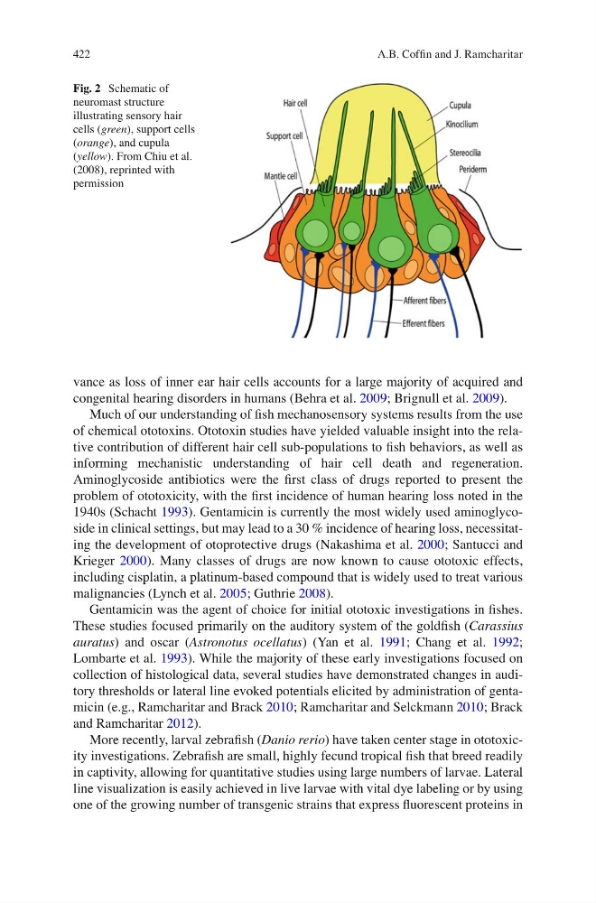
Fig. 2 Schematic of
neuromast structure
illustrating sensory hair
cells ( green ), support cells
( orange ), and cupula
( yellow ). From Chiu et al.
(2008), reprinted with
permission
A.B. Coffi n and J. Ramcharitar
vance as loss of inner ear hair cells accounts for a large majority of acquired and
congenital hearing disorders in humans (Behra et al. 2009 ; Brignull et al. 2009 ).
Much of our understanding of fi sh mechanosensory systems results from the use
of chemical ototoxins. Ototoxin studies have yielded valuable insight into the relative contribution of different hair cell sub-populations to fi sh behaviors, as well as
informing mechanistic understanding of hair cell death and regeneration.
Aminoglycoside antibiotics were the fi rst class of drugs reported to present the
problem of ototoxicity, with the fi rst incidence of human hearing loss noted in the
1940s (Schacht 1993 ). Gentamicin is currently the most widely used aminoglycoside in clinical settings, but may lead to a 30 % incidence of hearing loss, necessitating the development of otoprotective drugs (Nakashima et al. 2000 ; Santucci and
Krieger 2000 ). Many classes of drugs are now known to cause ototoxic effects,
including cisplatin, a platinum-based compound that is widely used to treat various
malignancies (Lynch et al. 2005 ; Guthrie 2008 ).
Gentamicin was the agent of choice for initial ototoxic investigations in fi shes.
These studies focused primarily on the auditory system of the goldfi sh ( Carassius
auratus ) and oscar ( Astronotus ocellatus ) (Yan et al. 1991 ; Chang et al. 1992 ;
Lombarte et al. 1993 ). While the majority of these early investigations focused on
collection of histological data, several studies have demonstrated changes in auditory thresholds or lateral line evoked potentials elicited by administration of gentamicin (e.g., Ramcharitar and Brack 2010 ; Ramcharitar and Selckmann 2010 ; Brack
and Ramcharitar 2012 ).
More recently, larval zebrafi sh ( Danio rerio ) have taken center stage in ototoxicity investigations. Zebrafi sh are small, highly fecund tropical fi sh that breed readily
in captivity, allowing for quantitative studies using large numbers of larvae. Lateral
line visualization is easily achieved in live larvae with vital dye labeling or by using
one of the growing number of transgenic strains that express fl uorescent proteins in
Fig. 2 Schematic of
neuromast structure
illustrating sensory hair
cells ( green ), support cells
( orange ), and cupula
( yellow ). From Chiu et al.
(2008), reprinted with
permission
A.B. Coffi n and J. Ramcharitar