

395
the trunk and tail (i.e., posterior lateral line) (Ghysen and Dambly-Chaudière 2004 ).
Adjacent to neuromasts are two types of accessory cells: support and mantle cells
(Villegas et al. 2012 ). Following hair cell damage, interior support cells proliferate
and differentiate into new hair cells (Ma et al. 2008 ). Neuromasts have cellular layers and nervous connections structurally similar to those found in the inner ear
sensory maculae (Nicolson 2005 ; Haehnel et al. 2012 ). Unlike inner ear hair cells,
lateral line stereociliary bundles project into a cupula, a gelatinous chamber, which
allows them to transduce vibrational forces into a neural signal (Nicolson 2005 ;
McHenry and van Netten 2007 ).
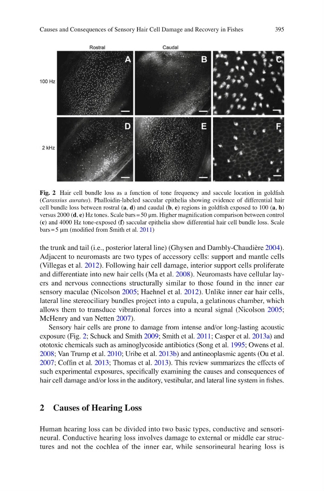
Sensory hair cells are prone to damage from intense and/or long-lasting acoustic
exposure (Fig. 2 ; Schuck and Smith 2009 ; Smith et al. 2011 ; Casper et al. 2013a ) and
ototoxic chemicals such as aminoglycoside antibiotics (Song et al. 1995 ; Owens et al.
2008 ; Van Trump et al. 2010 ; Uribe et al. 2013b ) and antineoplasmic agents (Ou et al.
2007 ; Coffi n et al. 2013 ; Thomas et al. 2013 ). This review summarizes the effects of
such experimental exposures, specifi cally examining the causes and consequences of
hair cell damage and/or loss in the auditory, vestibular, and lateral line system in fi shes.
2 Causes of Hearing Loss
Human hearing loss can be divided into two basic types, conductive and sensorineural. Conductive hearing loss involves damage to external or middle ear structures and not the cochlea of the inner ear, while sensorineural hearing loss is
Fig. 2 Hair cell bundle loss as a function of tone frequency and saccule location in goldfi sh
( Carassius auratus ). Phalloidin-labeled saccular epithelia showing evidence of differential hair
cell bundle loss between rostral ( a , d ) and caudal ( b , e ) regions in goldfi sh exposed to 100 ( a , b )
versus 2000 ( d , e ) Hz tones. Scale bars = 50 μm. Higher magnifi cation comparison between control
( c ) and 4000 Hz tone-exposed ( f ) saccular epithelia show differential hair cell bundle loss. Scale
bars = 5 μm (modifi ed from Smith et al. 2011 )
Causes and Consequences of Sensory Hair Cell Damage and Recovery in Fishes
the trunk and tail (i.e., posterior lateral line) (Ghysen and Dambly-Chaudière 2004 ).
Adjacent to neuromasts are two types of accessory cells: support and mantle cells
(Villegas et al. 2012 ). Following hair cell damage, interior support cells proliferate
and differentiate into new hair cells (Ma et al. 2008 ). Neuromasts have cellular layers and nervous connections structurally similar to those found in the inner ear
sensory maculae (Nicolson 2005 ; Haehnel et al. 2012 ). Unlike inner ear hair cells,
lateral line stereociliary bundles project into a cupula, a gelatinous chamber, which
allows them to transduce vibrational forces into a neural signal (Nicolson 2005 ;
McHenry and van Netten 2007 ).
Sensory hair cells are prone to damage from intense and/or long-lasting acoustic
exposure (Fig. 2 ; Schuck and Smith 2009 ; Smith et al. 2011 ; Casper et al. 2013a ) and
ototoxic chemicals such as aminoglycoside antibiotics (Song et al. 1995 ; Owens et al.
2008 ; Van Trump et al. 2010 ; Uribe et al. 2013b ) and antineoplasmic agents (Ou et al.
2007 ; Coffi n et al. 2013 ; Thomas et al. 2013 ). This review summarizes the effects of
such experimental exposures, specifi cally examining the causes and consequences of
hair cell damage and/or loss in the auditory, vestibular, and lateral line system in fi shes.
2 Causes of Hearing Loss
Human hearing loss can be divided into two basic types, conductive and sensorineural. Conductive hearing loss involves damage to external or middle ear structures and not the cochlea of the inner ear, while sensorineural hearing loss is
Fig. 2 Hair cell bundle loss as a function of tone frequency and saccule location in goldfi sh
( Carassius auratus ). Phalloidin-labeled saccular epithelia showing evidence of differential hair
cell bundle loss between rostral ( a , d ) and caudal ( b , e ) regions in goldfi sh exposed to 100 ( a , b )
versus 2000 ( d , e ) Hz tones. Scale bars = 50 μm. Higher magnifi cation comparison between control
( c ) and 4000 Hz tone-exposed ( f ) saccular epithelia show differential hair cell bundle loss. Scale
bars = 5 μm (modifi ed from Smith et al. 2011 )
Causes and Consequences of Sensory Hair Cell Damage and Recovery in Fishes