

394
acoustic overstimulation. In the latter, fi shes are exposed to acoustical stimuli from
man- made sources, such as seismic airguns, pile driving, sonars, and boat noise, or
signals that mimic these sound sources.
Although fi shes have no external or middle ears, their inner ears are similar to
those of other vertebrates. In fact, it is hypothesized that hearing fi rst evolved in
fi shes (Popper and Fay 1999 ). The fi sh ear consists of three semicircular canals
(except in more primitive taxa, i.e., hagfi shes and lampreys, which have fewer) and
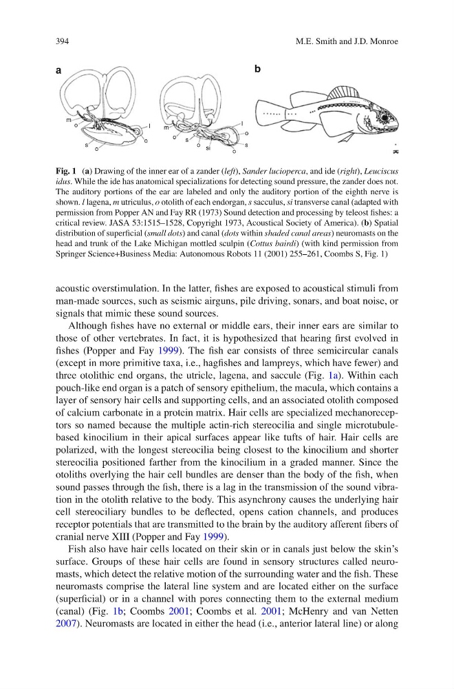
three otolithic end organs, the utricle, lagena, and saccule (Fig. 1a ). Within each
pouch-like end organ is a patch of sensory epithelium, the macula, which contains a
layer of sensory hair cells and supporting cells, and an associated otolith composed
of calcium carbonate in a protein matrix. Hair cells are specialized mechanoreceptors so named because the multiple actin-rich stereocilia and single microtubulebased kinocilium in their apical surfaces appear like tufts of hair. Hair cells are
polarized, with the longest stereocilia being closest to the kinocilium and shorter
stereocilia positioned farther from the kinocilium in a graded manner. Since the
otoliths overlying the hair cell bundles are denser than the body of the fi sh, when
sound passes through the fi sh, there is a lag in the transmission of the sound vibration in the otolith relative to the body. This asynchrony causes the underlying hair
cell stereociliary bundles to be defl ected, opens cation channels, and produces
receptor potentials that are transmitted to the brain by the auditory afferent fi bers of
cranial nerve XIII (Popper and Fay 1999 ).
Fish also have hair cells located on their skin or in canals just below the skin’s
surface. Groups of these hair cells are found in sensory structures called neuromasts, which detect the relative motion of the surrounding water and the fi sh. These
neuromasts comprise the lateral line system and are located either on the surface
(superfi cial) or in a channel with pores connecting them to the external medium
(canal) (Fig. 1b ; Coombs 2001 ; Coombs et al. 2001 ; McHenry and van Netten
2007 ). Neuromasts are located in either the head (i.e., anterior lateral line) or along
m
m
a
b
o
o
s
s
s
si
I
I
o
o
o
o
o
Fig. 1 ( a ) Drawing of the inner ear of a zander ( left ), Sander lucioperca , and ide ( right ), Leuciscus
idus . While the ide has anatomical specializations for detecting sound pressure, the zander does not.
The auditory portions of the ear are labeled and only the auditory portion of the eighth nerve is
shown. l lagena, m utriculus, o otolith of each endorgan, s sacculus, si transverse canal (adapted with
permission from Popper AN and Fay RR (1973) Sound detection and processing by teleost fi shes: a
critical review. JASA 53:1515–1528, Copyright 1973, Acoustical Society of America). ( b ) Spatial
distribution of superfi cial ( small dots ) and canal ( dots within shaded canal areas ) neuromasts on the
head and trunk of the Lake Michigan mottled sculpin ( Cottus bairdi ) (with kind permission from
Springer Science+Business Media: Autonomous Robots 11 (2001) 255–261, Coombs S, Fig. 1)
M.E. Smith and J.D. Monroe
acoustic overstimulation. In the latter, fi shes are exposed to acoustical stimuli from
man- made sources, such as seismic airguns, pile driving, sonars, and boat noise, or
signals that mimic these sound sources.
Although fi shes have no external or middle ears, their inner ears are similar to
those of other vertebrates. In fact, it is hypothesized that hearing fi rst evolved in
fi shes (Popper and Fay 1999 ). The fi sh ear consists of three semicircular canals
(except in more primitive taxa, i.e., hagfi shes and lampreys, which have fewer) and
three otolithic end organs, the utricle, lagena, and saccule (Fig. 1a ). Within each
pouch-like end organ is a patch of sensory epithelium, the macula, which contains a
layer of sensory hair cells and supporting cells, and an associated otolith composed
of calcium carbonate in a protein matrix. Hair cells are specialized mechanoreceptors so named because the multiple actin-rich stereocilia and single microtubulebased kinocilium in their apical surfaces appear like tufts of hair. Hair cells are
polarized, with the longest stereocilia being closest to the kinocilium and shorter
stereocilia positioned farther from the kinocilium in a graded manner. Since the
otoliths overlying the hair cell bundles are denser than the body of the fi sh, when
sound passes through the fi sh, there is a lag in the transmission of the sound vibration in the otolith relative to the body. This asynchrony causes the underlying hair
cell stereociliary bundles to be defl ected, opens cation channels, and produces
receptor potentials that are transmitted to the brain by the auditory afferent fi bers of
cranial nerve XIII (Popper and Fay 1999 ).
Fish also have hair cells located on their skin or in canals just below the skin’s
surface. Groups of these hair cells are found in sensory structures called neuromasts, which detect the relative motion of the surrounding water and the fi sh. These
neuromasts comprise the lateral line system and are located either on the surface
(superfi cial) or in a channel with pores connecting them to the external medium
(canal) (Fig. 1b ; Coombs 2001 ; Coombs et al. 2001 ; McHenry and van Netten
2007 ). Neuromasts are located in either the head (i.e., anterior lateral line) or along
m
m
a
b
o
o
s
s
s
si
I
I
o
o
o
o
o
Fig. 1 ( a ) Drawing of the inner ear of a zander ( left ), Sander lucioperca , and ide ( right ), Leuciscus
idus . While the ide has anatomical specializations for detecting sound pressure, the zander does not.
The auditory portions of the ear are labeled and only the auditory portion of the eighth nerve is
shown. l lagena, m utriculus, o otolith of each endorgan, s sacculus, si transverse canal (adapted with
permission from Popper AN and Fay RR (1973) Sound detection and processing by teleost fi shes: a
critical review. JASA 53:1515–1528, Copyright 1973, Acoustical Society of America). ( b ) Spatial
distribution of superfi cial ( small dots ) and canal ( dots within shaded canal areas ) neuromasts on the
head and trunk of the Lake Michigan mottled sculpin ( Cottus bairdi ) (with kind permission from
Springer Science+Business Media: Autonomous Robots 11 (2001) 255–261, Coombs S, Fig. 1)
M.E. Smith and J.D. Monroe