

233
2.2 Saccular Potential Recordings
Evoked saccular potentials from the Hawaiian sergeant fi sh were recorded at the
University of Washington. Adult A. abdominalis were caught as described above,
packaged individually in large bags fi lled with seawater and oxygen, and transported via overnight air-service to the Department of Psychology at the University
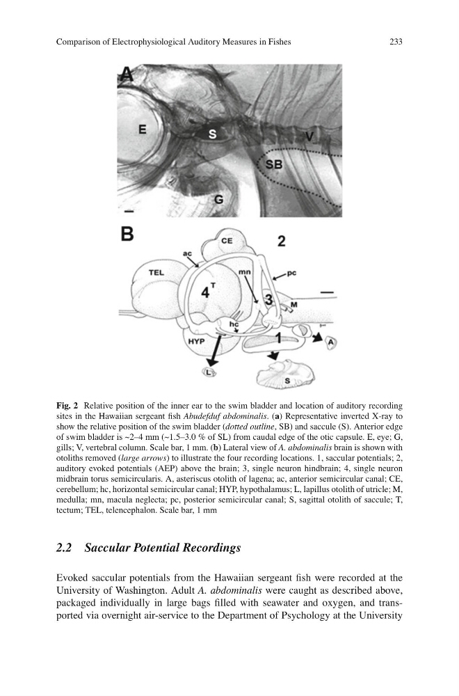
Fig. 2 Relative position of the inner ear to the swim bladder and location of auditory recording
sites in the Hawaiian sergeant fi sh Abudefduf abdominalis . ( a ) Representative inverted X-ray to
show the relative position of the swim bladder ( dotted outline , SB) and saccule (S). Anterior edge
of swim bladder is ~2–4 mm (~1.5–3.0 % of SL) from caudal edge of the otic capsule. E, eye; G,
gills; V, vertebral column. Scale bar, 1 mm. ( b ) Lateral view of A. abdominalis brain is shown with
otoliths removed ( large arrows ) to illustrate the four recording locations. 1, saccular potentials; 2,
auditory evoked potentials (AEP) above the brain; 3, single neuron hindbrain; 4, single neuron
midbrain torus semicircularis. A, asteriscus otolith of lagena; ac, anterior semicircular canal; CE,
cerebellum; hc, horizontal semicircular canal; HYP, hypothalamus; L, lapillus otolith of utricle; M,
medulla; mn, macula neglecta; pc, posterior semicircular canal; S, sagittal otolith of saccule; T,
tectum; TEL, telencephalon. Scale bar, 1 mm
Comparison of Electrophysiological Auditory Measures in Fishes
2.2 Saccular Potential Recordings
Evoked saccular potentials from the Hawaiian sergeant fi sh were recorded at the
University of Washington. Adult A. abdominalis were caught as described above,
packaged individually in large bags fi lled with seawater and oxygen, and transported via overnight air-service to the Department of Psychology at the University
Fig. 2 Relative position of the inner ear to the swim bladder and location of auditory recording
sites in the Hawaiian sergeant fi sh Abudefduf abdominalis . ( a ) Representative inverted X-ray to
show the relative position of the swim bladder ( dotted outline , SB) and saccule (S). Anterior edge
of swim bladder is ~2–4 mm (~1.5–3.0 % of SL) from caudal edge of the otic capsule. E, eye; G,
gills; V, vertebral column. Scale bar, 1 mm. ( b ) Lateral view of A. abdominalis brain is shown with
otoliths removed ( large arrows ) to illustrate the four recording locations. 1, saccular potentials; 2,
auditory evoked potentials (AEP) above the brain; 3, single neuron hindbrain; 4, single neuron
midbrain torus semicircularis. A, asteriscus otolith of lagena; ac, anterior semicircular canal; CE,
cerebellum; hc, horizontal semicircular canal; HYP, hypothalamus; L, lapillus otolith of utricle; M,
medulla; mn, macula neglecta; pc, posterior semicircular canal; S, sagittal otolith of saccule; T,
tectum; TEL, telencephalon. Scale bar, 1 mm
Comparison of Electrophysiological Auditory Measures in Fishes