

178
persistence of populations. However, in conservation, the application of molecular
markers can also contribute to the understanding of evolutionary history, demography and ecology of endangered species (Vilaça et al. 2016).
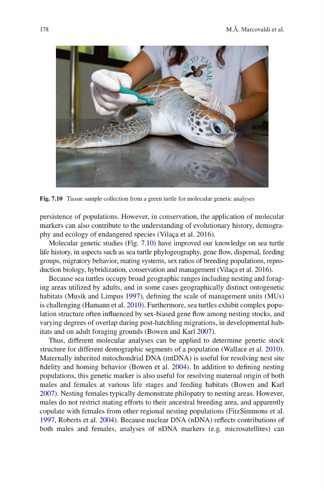
Molecular genetic studies (Fig. 7.10) have improved our knowledge on sea turtle
life history, in aspects such as sea turtle phylogeography, gene flow, dispersal, feeding
groups, migratory behavior, mating systems, sex ratios of breeding populations, reproduction biology, hybridization, conservation and management (Vilaça et al. 2016).
Because sea turtles occupy broad geographic ranges including nesting and foraging areas utilized by adults, and in some cases geographically distinct ontogenetic
habitats (Musik and Limpus 1997), defining the scale of management units (MUs)
is challenging (Hamann et al. 2010). Furthermore, sea turtles exhibit complex population structure often influenced by sex-biased gene flow among nesting stocks, and
varying degrees of overlap during post-hatchling migrations, in developmental habitats and on adult foraging grounds (Bowen and Karl 2007).
Thus, different molecular analyses can be applied to determine genetic stock
structure for different demographic segments of a population (Wallace et al. 2010).
Maternally inherited mitochondrial DNA (mtDNA) is useful for resolving nest site
fidelity and homing behavior (Bowen et al. 2004). In addition to defining nesting
populations, this genetic marker is also useful for resolving maternal origin of both
males and females at various life stages and feeding habitats (Bowen and Karl
2007). Nesting females typically demonstrate philopatry to nesting areas. However,
males do not restrict mating efforts to their ancestral breeding area, and apparently
copulate with females from other regional nesting populations (FitzSimmons et al.
1997, Roberts et al. 2004). Because nuclear DNA (nDNA) reflects contributions of
both males and females, analyses of nDNA markers (e.g. microsatellites) can
Fig. 7.10 Tissue sample collection from a green turtle for molecular genetic analyses
M.Â. Marcovaldi et al.
persistence of populations. However, in conservation, the application of molecular
markers can also contribute to the understanding of evolutionary history, demography and ecology of endangered species (Vilaça et al. 2016).
Molecular genetic studies (Fig. 7.10) have improved our knowledge on sea turtle
life history, in aspects such as sea turtle phylogeography, gene flow, dispersal, feeding
groups, migratory behavior, mating systems, sex ratios of breeding populations, reproduction biology, hybridization, conservation and management (Vilaça et al. 2016).
Because sea turtles occupy broad geographic ranges including nesting and foraging areas utilized by adults, and in some cases geographically distinct ontogenetic
habitats (Musik and Limpus 1997), defining the scale of management units (MUs)
is challenging (Hamann et al. 2010). Furthermore, sea turtles exhibit complex population structure often influenced by sex-biased gene flow among nesting stocks, and
varying degrees of overlap during post-hatchling migrations, in developmental habitats and on adult foraging grounds (Bowen and Karl 2007).
Thus, different molecular analyses can be applied to determine genetic stock
structure for different demographic segments of a population (Wallace et al. 2010).
Maternally inherited mitochondrial DNA (mtDNA) is useful for resolving nest site
fidelity and homing behavior (Bowen et al. 2004). In addition to defining nesting
populations, this genetic marker is also useful for resolving maternal origin of both
males and females at various life stages and feeding habitats (Bowen and Karl
2007). Nesting females typically demonstrate philopatry to nesting areas. However,
males do not restrict mating efforts to their ancestral breeding area, and apparently
copulate with females from other regional nesting populations (FitzSimmons et al.
1997, Roberts et al. 2004). Because nuclear DNA (nDNA) reflects contributions of
both males and females, analyses of nDNA markers (e.g. microsatellites) can
Fig. 7.10 Tissue sample collection from a green turtle for molecular genetic analyses
M.Â. Marcovaldi et al.