

chain reaction (PCR) assay is available as a diagnostic tool
and a fluorescent in situ hybridization (FISH) has the potential
detect F. psychrophilum in infected tissues. Flavobacterium
psychrophilum exists in at least three serotypes with recognised variation in their virulence.
6.9
Flavobacterium columnare
Flavobacterium columnare, formerly known as Flexibacter
columnaris, is the causative agent of ‘columnaris disease’,
a bacterial infection affecting freshwater fish including rainbow trout. Outbreaks are related to stress including high
temperature, low levels of dissolved oxygen, increased
ammonia and organic load. Infection is transmitted
horizontally through direct contact with infected fish.
Early clinical signs are nonspecific and include lethargy,
inappetence, and swimming near the water surface with
increased opercula movements. Additional and more characteristic signs include skin discoloration, dorsal fin damage
(Fig. 6.26) and yellow necrotic gill lesions at the tips of
the lamellae. As lesions progress to the mandible and maxillae
the paucity of underlying tissue lesion leads to fatal osmotic
changes. In addition, hypoxia caused by gill necrosis and
biochemical disturbances resulting from skin ulceration, are
a likely cause of death. Bacterial attachment to the gill tissue
and entry into the fish is facilitated by physical injuries thus
highlighting the importance of the gills in pathogenesis. Internally, ascites can be observed.
Histologically, lamellar shortening, epithelial and goblet
cell hyperplasia with an associated moderate necrosis is
reported. These lesions rapidly progress to a marked inflammation predominantly by neutrophils and severe gill necrosis. In acute infections, hypoxia and death may result from
extensive damage to this tissue.
Diagnosis is based upon characteristic clinical signs and
histopathology, and the isolation and identification of the
bacteria. Flavobacterium columnare is a Gram negative thin
rod-shaped bacterium which measures 3–5 μm in length.
Isolation of the bacteria can be made from the gill, surface
lesions as well as internal organs such as the kidney, liver and
spleen. In wet mounts bacteria are uniquely arranged into
columnar formations. A definitive diagnosis of F.columnare
is dependent upon culture using a selective medium
(e.g. Cytophaga or Shieh) with the presence of yellow
pigmented rhizoid colonies. This is followed by biochemical
testing using conventional methods.
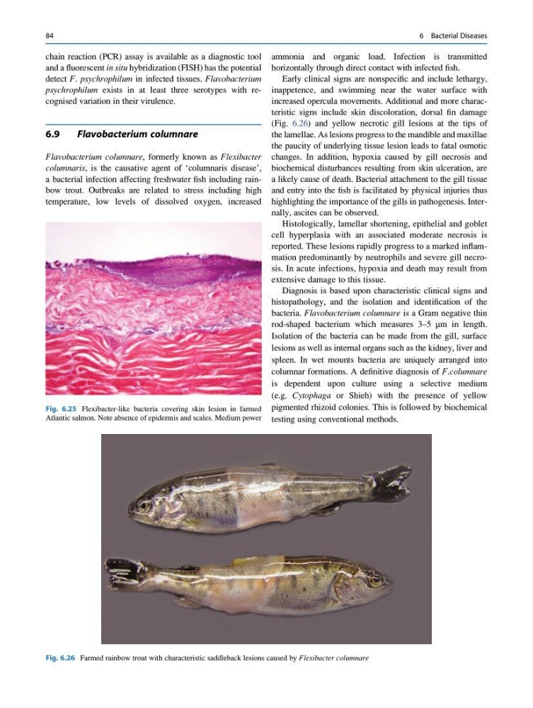
Fig. 6.25 Flexibacter-like bacteria covering skin lesion in farmed
Atlantic salmon. Note absence of epidermis and scales. Medium power
Fig. 6.26 Farmed rainbow trout with characteristic saddleback lesions caused by Flexibacter columnare
84
6 Bacterial Diseases
and a fluorescent in situ hybridization (FISH) has the potential
detect F. psychrophilum in infected tissues. Flavobacterium
psychrophilum exists in at least three serotypes with recognised variation in their virulence.
6.9
Flavobacterium columnare
Flavobacterium columnare, formerly known as Flexibacter
columnaris, is the causative agent of ‘columnaris disease’,
a bacterial infection affecting freshwater fish including rainbow trout. Outbreaks are related to stress including high
temperature, low levels of dissolved oxygen, increased
ammonia and organic load. Infection is transmitted
horizontally through direct contact with infected fish.
Early clinical signs are nonspecific and include lethargy,
inappetence, and swimming near the water surface with
increased opercula movements. Additional and more characteristic signs include skin discoloration, dorsal fin damage
(Fig. 6.26) and yellow necrotic gill lesions at the tips of
the lamellae. As lesions progress to the mandible and maxillae
the paucity of underlying tissue lesion leads to fatal osmotic
changes. In addition, hypoxia caused by gill necrosis and
biochemical disturbances resulting from skin ulceration, are
a likely cause of death. Bacterial attachment to the gill tissue
and entry into the fish is facilitated by physical injuries thus
highlighting the importance of the gills in pathogenesis. Internally, ascites can be observed.
Histologically, lamellar shortening, epithelial and goblet
cell hyperplasia with an associated moderate necrosis is
reported. These lesions rapidly progress to a marked inflammation predominantly by neutrophils and severe gill necrosis. In acute infections, hypoxia and death may result from
extensive damage to this tissue.
Diagnosis is based upon characteristic clinical signs and
histopathology, and the isolation and identification of the
bacteria. Flavobacterium columnare is a Gram negative thin
rod-shaped bacterium which measures 3–5 μm in length.
Isolation of the bacteria can be made from the gill, surface
lesions as well as internal organs such as the kidney, liver and
spleen. In wet mounts bacteria are uniquely arranged into
columnar formations. A definitive diagnosis of F.columnare
is dependent upon culture using a selective medium
(e.g. Cytophaga or Shieh) with the presence of yellow
pigmented rhizoid colonies. This is followed by biochemical
testing using conventional methods.
Fig. 6.25 Flexibacter-like bacteria covering skin lesion in farmed
Atlantic salmon. Note absence of epidermis and scales. Medium power
Fig. 6.26 Farmed rainbow trout with characteristic saddleback lesions caused by Flexibacter columnare
84
6 Bacterial Diseases