

based upon clinical signs, and the isolation and identification
of the bacteria. A differential diagnosis would include other
Vibrio spp.
Moritella viscosa is a psychrophilic Gram negative, motile,
flagellated curved rod. M. viscosa antigens are included in
most of the multivalent injection vaccines used in the salmon
industry, but the protection is apparently variable.
6.6
Yersinia ruckeri
Yersinia ruckeri is the causative agent of yersiniosis or enteric
redmouth disease (ERM), an economically important condition in wild and farmed salmonids, both in fresh and sea
water. Y. ruckeri has a wide host range and most salmonids
are susceptible. Transmission occurs horizontally and many
species of asymptomatic carriers as well as birds, being
reservoirs of infection. Outbreaks are typically stressmediated, e.g. poor water quality, increase in temperature,
grading and handling. Clinical signs and pathology are similar
to other Gram negative septicaemias and the disease may
occur in peracute to acute or more chronic forms. In peracute
cases, as in fry or fingerlings in freshwater, there may be high
mortality with few or no external disease signs. In the more
chronic cases, fish show pigment changes, disturbance of
balance and lethargy. Other signs include ascites, exophthalmia, cutaneous petechiae and localized haemorrhage
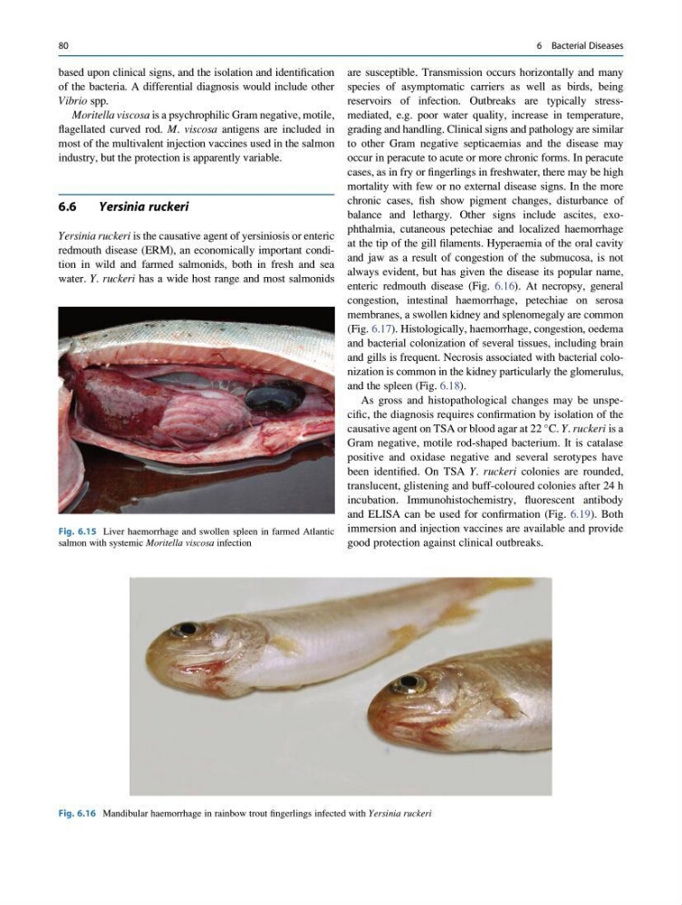
at the tip of the gill filaments. Hyperaemia of the oral cavity
and jaw as a result of congestion of the submucosa, is not
always evident, but has given the disease its popular name,
enteric redmouth disease (Fig. 6.16). At necropsy, general
congestion, intestinal haemorrhage, petechiae on serosa
membranes, a swollen kidney and splenomegaly are common
(Fig. 6.17). Histologically, haemorrhage, congestion, oedema
and bacterial colonization of several tissues, including brain
and gills is frequent. Necrosis associated with bacterial colonization is common in the kidney particularly the glomerulus,
and the spleen (Fig. 6.18).
As gross and histopathological changes may be unspecific, the diagnosis requires confirmation by isolation of the
causative agent on TSA or blood agar at 22
C. Y. ruckeri is a
Gram negative, motile rod-shaped bacterium. It is catalase
positive and oxidase negative and several serotypes have
been identified. On TSA Y. ruckeri colonies are rounded,
translucent, glistening and buff-coloured colonies after 24 h
incubation. Immunohistochemistry, fluorescent antibody
and ELISA can be used for confirmation (Fig. 6.19). Both
immersion and injection vaccines are available and provide
good protection against clinical outbreaks.
Fig. 6.15 Liver haemorrhage and swollen spleen in farmed Atlantic
salmon with systemic Moritella viscosa infection
Fig. 6.16 Mandibular haemorrhage in rainbow trout fingerlings infected with Yersinia ruckeri
80
6 Bacterial Diseases
of the bacteria. A differential diagnosis would include other
Vibrio spp.
Moritella viscosa is a psychrophilic Gram negative, motile,
flagellated curved rod. M. viscosa antigens are included in
most of the multivalent injection vaccines used in the salmon
industry, but the protection is apparently variable.
6.6
Yersinia ruckeri
Yersinia ruckeri is the causative agent of yersiniosis or enteric
redmouth disease (ERM), an economically important condition in wild and farmed salmonids, both in fresh and sea
water. Y. ruckeri has a wide host range and most salmonids
are susceptible. Transmission occurs horizontally and many
species of asymptomatic carriers as well as birds, being
reservoirs of infection. Outbreaks are typically stressmediated, e.g. poor water quality, increase in temperature,
grading and handling. Clinical signs and pathology are similar
to other Gram negative septicaemias and the disease may
occur in peracute to acute or more chronic forms. In peracute
cases, as in fry or fingerlings in freshwater, there may be high
mortality with few or no external disease signs. In the more
chronic cases, fish show pigment changes, disturbance of
balance and lethargy. Other signs include ascites, exophthalmia, cutaneous petechiae and localized haemorrhage
at the tip of the gill filaments. Hyperaemia of the oral cavity
and jaw as a result of congestion of the submucosa, is not
always evident, but has given the disease its popular name,
enteric redmouth disease (Fig. 6.16). At necropsy, general
congestion, intestinal haemorrhage, petechiae on serosa
membranes, a swollen kidney and splenomegaly are common
(Fig. 6.17). Histologically, haemorrhage, congestion, oedema
and bacterial colonization of several tissues, including brain
and gills is frequent. Necrosis associated with bacterial colonization is common in the kidney particularly the glomerulus,
and the spleen (Fig. 6.18).
As gross and histopathological changes may be unspecific, the diagnosis requires confirmation by isolation of the
causative agent on TSA or blood agar at 22
C. Y. ruckeri is a
Gram negative, motile rod-shaped bacterium. It is catalase
positive and oxidase negative and several serotypes have
been identified. On TSA Y. ruckeri colonies are rounded,
translucent, glistening and buff-coloured colonies after 24 h
incubation. Immunohistochemistry, fluorescent antibody
and ELISA can be used for confirmation (Fig. 6.19). Both
immersion and injection vaccines are available and provide
good protection against clinical outbreaks.
Fig. 6.15 Liver haemorrhage and swollen spleen in farmed Atlantic
salmon with systemic Moritella viscosa infection
Fig. 6.16 Mandibular haemorrhage in rainbow trout fingerlings infected with Yersinia ruckeri
80
6 Bacterial Diseases