

extending from the outer compact layer into the spongy layer
with slight loss of striation. Lesions progress from focal to
multifocal or diffuse degeneration of the trabecular myocardium and increased number of mononuclear inflammatory
cells, lymphocytes and plasma cells, around multiple intramural coronary vessels and, intermittently, throughout the
epicardium (Figs. 5.41 and 5.42). These cells frequently
invade the sub-endocardial spaces (Fig. 5.43). Associated
lesions may include mural thrombi (Fig. 5.44), hypertrophy
of myocardial nuclei, which are believed to represent a
compensatory reaction in a failing heart, and ‘nest-like’
aggregates of nuclei. The compact ventricular myocardium
is usually unaffected, but a highly cellular epicarditis is
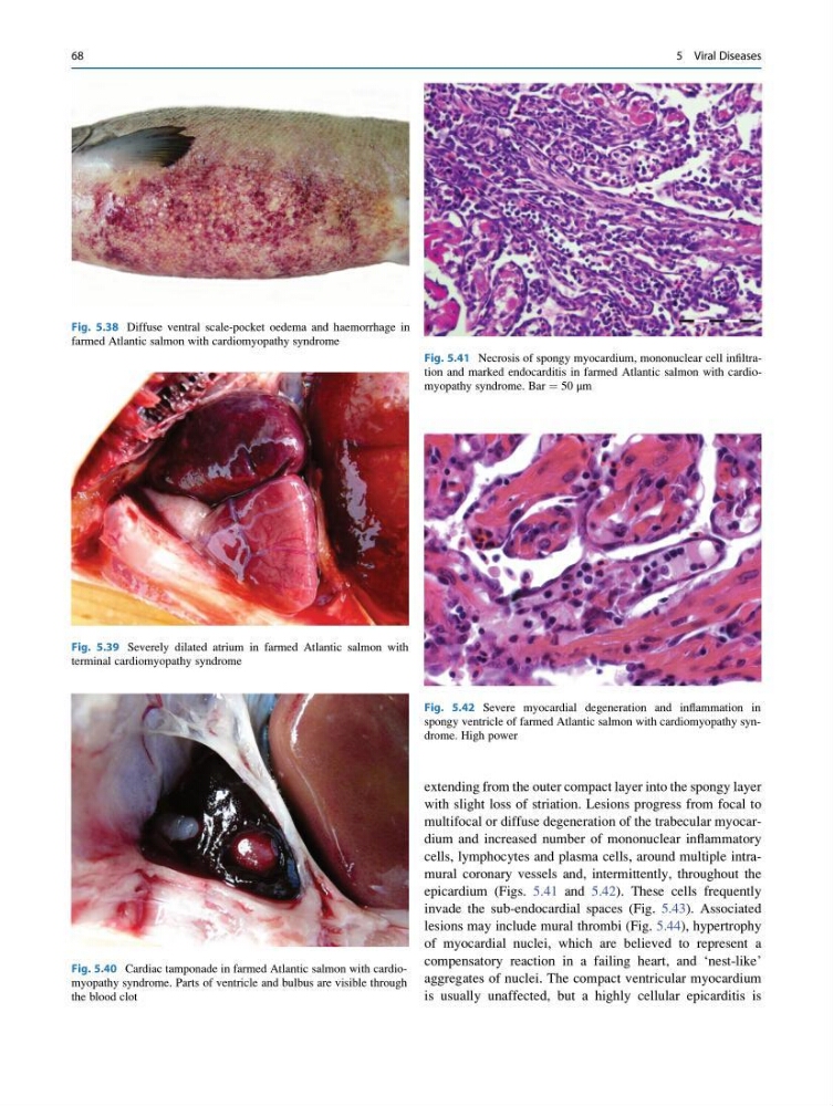
Fig. 5.39 Severely dilated atrium in farmed Atlantic salmon with
terminal cardiomyopathy syndrome
Fig. 5.40 Cardiac tamponade in farmed Atlantic salmon with cardiomyopathy syndrome. Parts of ventricle and bulbus are visible through
the blood clot
Fig. 5.41 Necrosis of spongy myocardium, mononuclear cell infiltration and marked endocarditis in farmed Atlantic salmon with cardiomyopathy syndrome. Bar ¼ 50 μm
Fig. 5.42 Severe myocardial degeneration and inflammation in
spongy ventricle of farmed Atlantic salmon with cardiomyopathy syndrome. High power
Fig. 5.38 Diffuse ventral scale-pocket oedema and haemorrhage in
farmed Atlantic salmon with cardiomyopathy syndrome
68
5 Viral Diseases
with slight loss of striation. Lesions progress from focal to
multifocal or diffuse degeneration of the trabecular myocardium and increased number of mononuclear inflammatory
cells, lymphocytes and plasma cells, around multiple intramural coronary vessels and, intermittently, throughout the
epicardium (Figs. 5.41 and 5.42). These cells frequently
invade the sub-endocardial spaces (Fig. 5.43). Associated
lesions may include mural thrombi (Fig. 5.44), hypertrophy
of myocardial nuclei, which are believed to represent a
compensatory reaction in a failing heart, and ‘nest-like’
aggregates of nuclei. The compact ventricular myocardium
is usually unaffected, but a highly cellular epicarditis is
Fig. 5.39 Severely dilated atrium in farmed Atlantic salmon with
terminal cardiomyopathy syndrome
Fig. 5.40 Cardiac tamponade in farmed Atlantic salmon with cardiomyopathy syndrome. Parts of ventricle and bulbus are visible through
the blood clot
Fig. 5.41 Necrosis of spongy myocardium, mononuclear cell infiltration and marked endocarditis in farmed Atlantic salmon with cardiomyopathy syndrome. Bar ¼ 50 μm
Fig. 5.42 Severe myocardial degeneration and inflammation in
spongy ventricle of farmed Atlantic salmon with cardiomyopathy syndrome. High power
Fig. 5.38 Diffuse ventral scale-pocket oedema and haemorrhage in
farmed Atlantic salmon with cardiomyopathy syndrome
68
5 Viral Diseases