

The diagnosis of PD is currently based on a combination
of histopathological examination, virus culture and PCR
detection using heart tissue samples rather than kidney.
The differential diagnosis includes CMS and HSMI. There
is evidence that outbreaks are declining and attributed to
improved management practices and the wider application
of available vaccines.
5.10 Piscine Myocarditis Virus
(Cardiomyopathy Syndrome)
Piscine myocarditis virus (PMV) has recently been recognised
as the aetiology of cardiomyopathy syndrome (CMS), a
chronic heart disease primarily affecting marine farmed Atlantic salmon. Lesions identical to those seen in fish with CMS
have also been recorded in wild salmon but the disease has not
been diagnosed in other salmonids. The first cases were
reported from Norway in 1985, but subsequently described
from other salmon-farming countries including Scotland, The
Faroe Islands and Canada. CMS is a chronic progressive
disease that develops over several months, with mortality
typically occurring in large fish 12–18 months after transfer
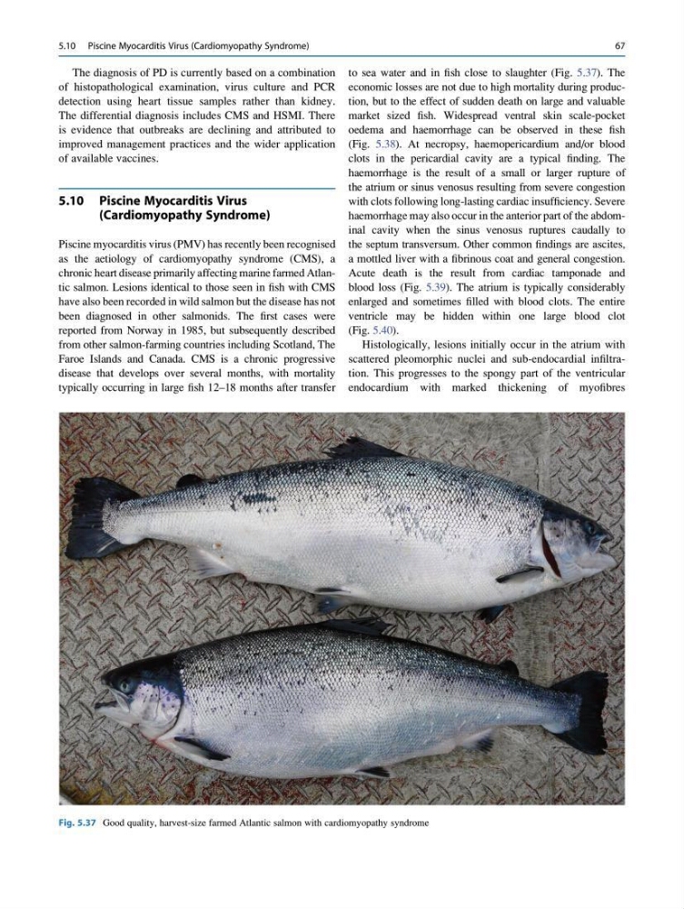
to sea water and in fish close to slaughter (Fig. 5.37). The
economic losses are not due to high mortality during production, but to the effect of sudden death on large and valuable
market sized fish. Widespread ventral skin scale-pocket
oedema and haemorrhage can be observed in these fish
(Fig. 5.38). At necropsy, haemopericardium and/or blood
clots in the pericardial cavity are a typical finding. The
haemorrhage is the result of a small or larger rupture of
the atrium or sinus venosus resulting from severe congestion
with clots following long-lasting cardiac insufficiency. Severe
haemorrhage may also occur in the anterior part of the abdominal cavity when the sinus venosus ruptures caudally to
the septum transversum. Other common findings are ascites,
a mottled liver with a fibrinous coat and general congestion.
Acute death is the result from cardiac tamponade and
blood loss (Fig. 5.39). The atrium is typically considerably
enlarged and sometimes filled with blood clots. The entire
ventricle may be hidden within one large blood clot
(Fig. 5.40).
Histologically, lesions initially occur in the atrium with
scattered pleomorphic nuclei and sub-endocardial infiltration. This progresses to the spongy part of the ventricular
endocardium with marked thickening of myofibres
Fig. 5.37 Good quality, harvest-size farmed Atlantic salmon with cardiomyopathy syndrome
5.10 Piscine Myocarditis Virus (Cardiomyopathy Syndrome)
67
of histopathological examination, virus culture and PCR
detection using heart tissue samples rather than kidney.
The differential diagnosis includes CMS and HSMI. There
is evidence that outbreaks are declining and attributed to
improved management practices and the wider application
of available vaccines.
5.10 Piscine Myocarditis Virus
(Cardiomyopathy Syndrome)
Piscine myocarditis virus (PMV) has recently been recognised
as the aetiology of cardiomyopathy syndrome (CMS), a
chronic heart disease primarily affecting marine farmed Atlantic salmon. Lesions identical to those seen in fish with CMS
have also been recorded in wild salmon but the disease has not
been diagnosed in other salmonids. The first cases were
reported from Norway in 1985, but subsequently described
from other salmon-farming countries including Scotland, The
Faroe Islands and Canada. CMS is a chronic progressive
disease that develops over several months, with mortality
typically occurring in large fish 12–18 months after transfer
to sea water and in fish close to slaughter (Fig. 5.37). The
economic losses are not due to high mortality during production, but to the effect of sudden death on large and valuable
market sized fish. Widespread ventral skin scale-pocket
oedema and haemorrhage can be observed in these fish
(Fig. 5.38). At necropsy, haemopericardium and/or blood
clots in the pericardial cavity are a typical finding. The
haemorrhage is the result of a small or larger rupture of
the atrium or sinus venosus resulting from severe congestion
with clots following long-lasting cardiac insufficiency. Severe
haemorrhage may also occur in the anterior part of the abdominal cavity when the sinus venosus ruptures caudally to
the septum transversum. Other common findings are ascites,
a mottled liver with a fibrinous coat and general congestion.
Acute death is the result from cardiac tamponade and
blood loss (Fig. 5.39). The atrium is typically considerably
enlarged and sometimes filled with blood clots. The entire
ventricle may be hidden within one large blood clot
(Fig. 5.40).
Histologically, lesions initially occur in the atrium with
scattered pleomorphic nuclei and sub-endocardial infiltration. This progresses to the spongy part of the ventricular
endocardium with marked thickening of myofibres
Fig. 5.37 Good quality, harvest-size farmed Atlantic salmon with cardiomyopathy syndrome
5.10 Piscine Myocarditis Virus (Cardiomyopathy Syndrome)
67