

2.10 Reproductive System
2.10.1 Ovary
The ovaries are paired sac-like organs in the dorsolateral part
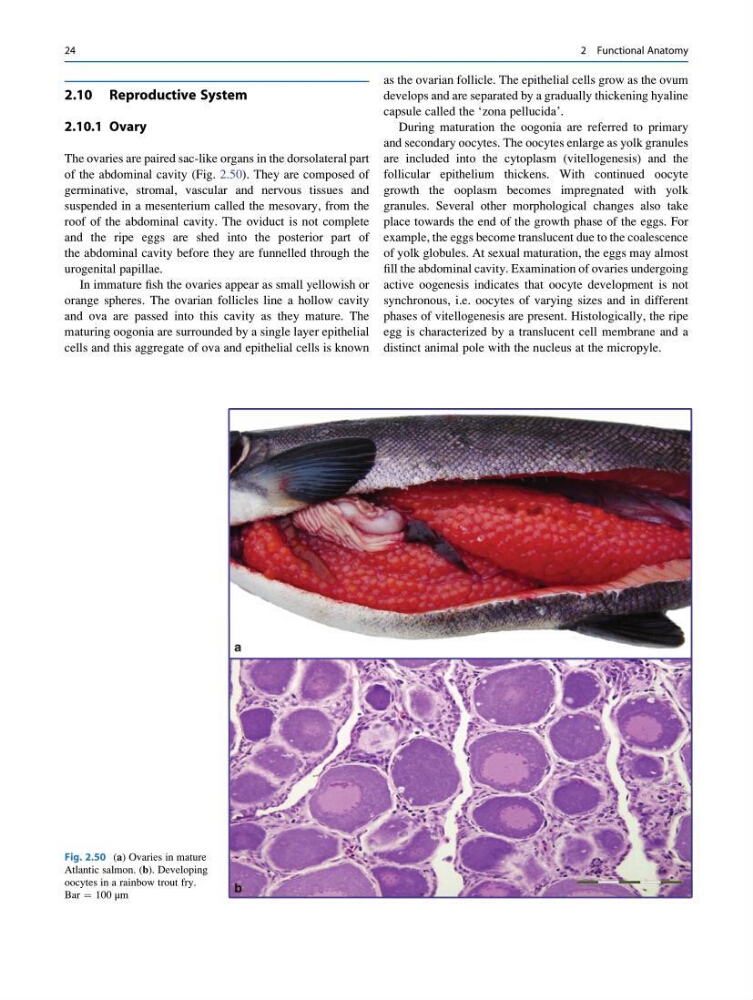
of the abdominal cavity (Fig. 2.50). They are composed of
germinative, stromal, vascular and nervous tissues and
suspended in a mesenterium called the mesovary, from the
roof of the abdominal cavity. The oviduct is not complete
and the ripe eggs are shed into the posterior part of
the abdominal cavity before they are funnelled through the
urogenital papillae.
In immature fish the ovaries appear as small yellowish or
orange spheres. The ovarian follicles line a hollow cavity
and ova are passed into this cavity as they mature. The
maturing oogonia are surrounded by a single layer epithelial
cells and this aggregate of ova and epithelial cells is known
as the ovarian follicle. The epithelial cells grow as the ovum
develops and are separated by a gradually thickening hyaline
capsule called the ‘zona pellucida’.
During maturation the oogonia are referred to primary
and secondary oocytes. The oocytes enlarge as yolk granules
are included into the cytoplasm (vitellogenesis) and the
follicular epithelium thickens. With continued oocyte
growth the ooplasm becomes impregnated with yolk
granules. Several other morphological changes also take
place towards the end of the growth phase of the eggs. For
example, the eggs become translucent due to the coalescence
of yolk globules. At sexual maturation, the eggs may almost
fill the abdominal cavity. Examination of ovaries undergoing
active oogenesis indicates that oocyte development is not
synchronous, i.e. oocytes of varying sizes and in different
phases of vitellogenesis are present. Histologically, the ripe
egg is characterized by a translucent cell membrane and a
distinct animal pole with the nucleus at the micropyle.
Fig. 2.50 (a) Ovaries in mature
Atlantic salmon. (b). Developing
oocytes in a rainbow trout fry.
Bar ¼ 100 μm
24
2 Functional Anatomy
2.10.1 Ovary
The ovaries are paired sac-like organs in the dorsolateral part
of the abdominal cavity (Fig. 2.50). They are composed of
germinative, stromal, vascular and nervous tissues and
suspended in a mesenterium called the mesovary, from the
roof of the abdominal cavity. The oviduct is not complete
and the ripe eggs are shed into the posterior part of
the abdominal cavity before they are funnelled through the
urogenital papillae.
In immature fish the ovaries appear as small yellowish or
orange spheres. The ovarian follicles line a hollow cavity
and ova are passed into this cavity as they mature. The
maturing oogonia are surrounded by a single layer epithelial
cells and this aggregate of ova and epithelial cells is known
as the ovarian follicle. The epithelial cells grow as the ovum
develops and are separated by a gradually thickening hyaline
capsule called the ‘zona pellucida’.
During maturation the oogonia are referred to primary
and secondary oocytes. The oocytes enlarge as yolk granules
are included into the cytoplasm (vitellogenesis) and the
follicular epithelium thickens. With continued oocyte
growth the ooplasm becomes impregnated with yolk
granules. Several other morphological changes also take
place towards the end of the growth phase of the eggs. For
example, the eggs become translucent due to the coalescence
of yolk globules. At sexual maturation, the eggs may almost
fill the abdominal cavity. Examination of ovaries undergoing
active oogenesis indicates that oocyte development is not
synchronous, i.e. oocytes of varying sizes and in different
phases of vitellogenesis are present. Histologically, the ripe
egg is characterized by a translucent cell membrane and a
distinct animal pole with the nucleus at the micropyle.
Fig. 2.50 (a) Ovaries in mature
Atlantic salmon. (b). Developing
oocytes in a rainbow trout fry.
Bar ¼ 100 μm
24
2 Functional Anatomy